
Cervical MRI Report Phrases Explained in Plain Words
What your neck MRI report means: uncovertebral hypertrophy, foraminal narrowing grades, effacement of the CSF, cord flattening, T2 hyperintense cord signal, Modic changes and more, each explained in one or two plain sentences.
Key Takeaways
- Reports describe each level in order: disc, central canal and cord, then the two nerve exits
- Uncovertebral hypertrophy is spurring unique to the neck and a common cause of foraminal narrowing
- Effacement, abutment, flattening and compression describe rising pressure on the cord
- T2 hyperintense cord signal with hand or walking symptoms needs prompt specialist review
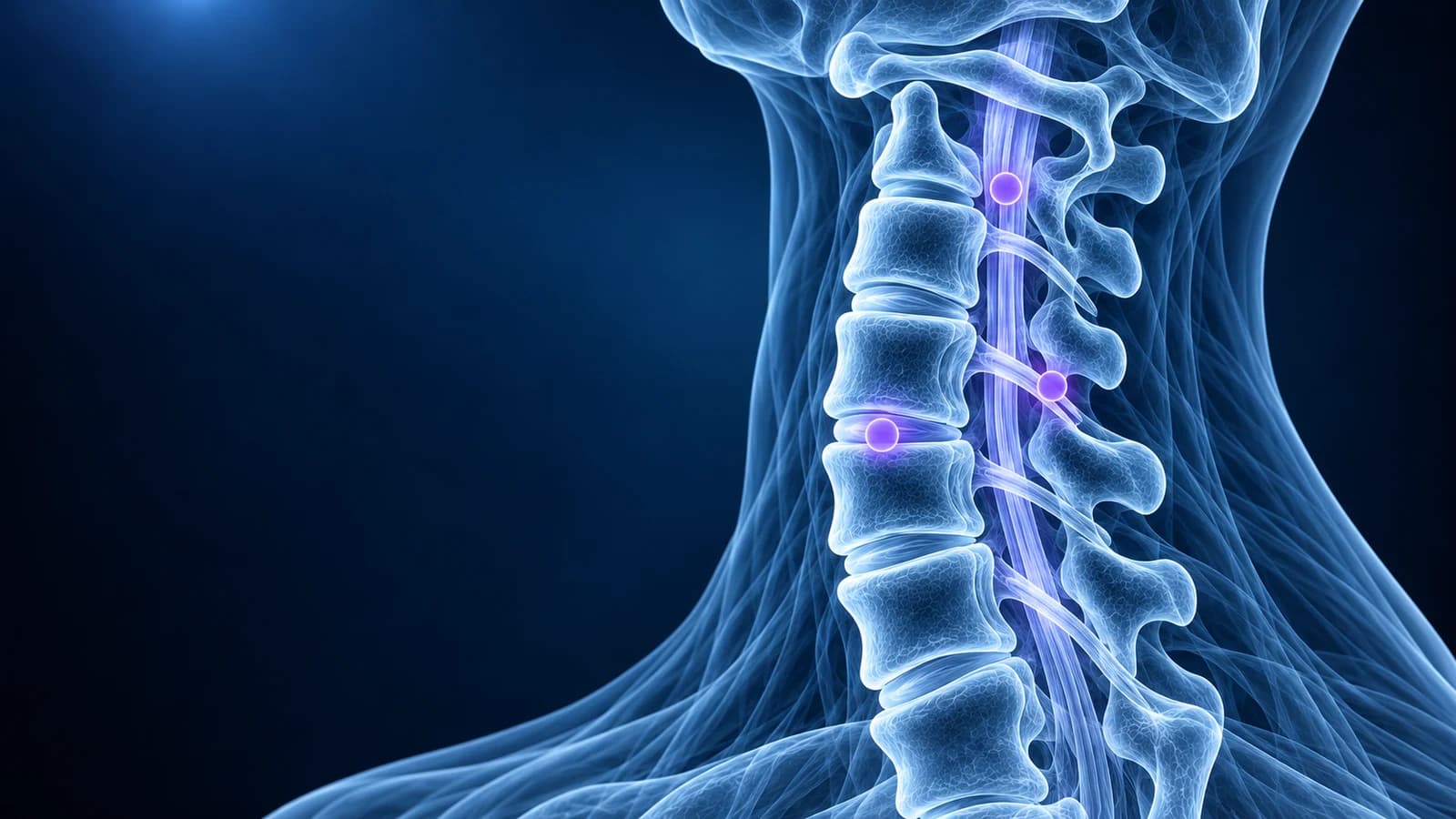
A cervical MRI report describes each disc level from C2-C3 to C7-T1 in the same order: the disc, the central canal around the spinal cord, and the two nerve exits (foramina). “Uncovertebral hypertrophy” means bone spurs at the side of a disc, “foraminal narrowing” means a tighter nerve exit, “effacement of the CSF” means fluid around the cord is pushed aside, and “T2 hyperintense cord signal” means a bright spot inside the cord, the phrase that most needs prompt follow-up.
Below, each phrase is explained in one or two sentences and linked to the condition it usually belongs to. Many of these findings are common with age and appear in people with no symptoms, so a phrase only matters when it matches your symptoms and examination. This page is educational, not a diagnosis.
What do the disc phrases in a neck MRI report mean?
- Disc desiccation: the disc has lost water and looks darker on T2 images. It is the earliest age change and on its own rarely explains pain.
- Loss of disc height: the disc is thinner than its neighbours, so the bones above and below sit closer together.
- Diffuse disc bulge: a broad, shallow overhang around much of the disc edge. Common from middle age and often seen without symptoms.
- Protrusion, extrusion, sequestration: increasingly focal disc herniations, from a short broad bump to material that has squeezed out or broken off. They are the hallmark of cervical disc herniation, most often at the two levels compared in C5-C6 and C6-C7 disc herniation explained.
- Central, paracentral, foraminal: where the disc points. Central toward the cord, paracentral slightly to one side, foraminal into the nerve exit.
- Annular fissure (sometimes “annular tear”): a small bright crack in the outer ring of the disc. It describes the disc’s structure and is not an injury in the usual sense.
- Disc osteophyte complex: a bulging disc and bone spurs from the vertebral edges merged into one hard ridge. Explained in detail in disc osteophyte complex explained.
What does “uncovertebral hypertrophy” mean?
The uncovertebral joints (joints of Luschka) are small lips of bone along the sides of each cervical disc from C3 to C7; they exist only in the neck. With age they thicken and grow spurs, which the report calls “uncovertebral hypertrophy”, “uncovertebral spurring” or “uncinate osteophytes”. Because they sit right at the front edge of the nerve exit, they are one of the main reasons a foramen narrows in the neck. Together with facet joint wear, they make up cervical spondylosis.
“Facet hypertrophy” or “facet arthropathy” is the same wear process in the small joints at the back of the spine. Facet joints narrow the foramen from behind, and arthritic facets at the top of the neck are one recognised source of cervicogenic headache.
What do mild, moderate and severe foraminal narrowing mean?
“Foraminal narrowing” or “neural foraminal stenosis” means the bony tunnel where a nerve root leaves the spine is smaller than normal, usually because of uncovertebral spurs, facet hypertrophy or a disc bulging sideways. Grading varies between radiologists, but a widely cited system based on oblique sagittal images works like this:
- Grade 0 (none): the fat around the nerve root is intact.
- Grade 1 (mild): fat is lost around less than half of the nerve root.
- Grade 2 (moderate): fat is lost around more than half of the root, but the root keeps its shape.
- Grade 3 (severe): the nerve root itself looks squeezed or collapsed.
The level tells you which root is involved: C5-C6 narrowing affects the C6 root and C6-C7 the C7 root. Narrowing on the side of your arm symptoms fits cervical foraminal stenosis, and if the root is irritated enough to cause pain, tingling or weakness down the arm, the clinical diagnosis is cervical radiculopathy. “Abuts”, “contacts” or “impinges on the exiting nerve root” describe the same relationship from mild to more definite.
What does “effacement of the CSF” or “cord flattening” mean?
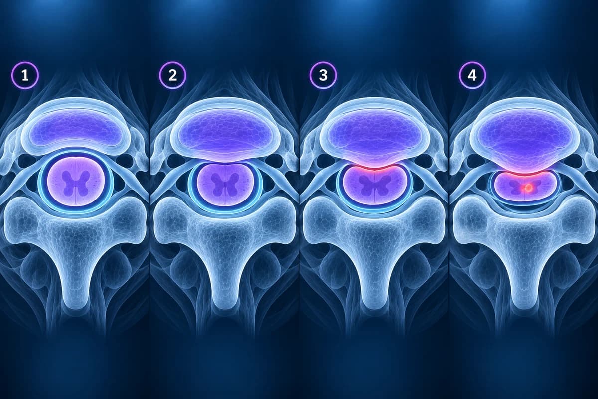
- Effacement of the ventral (or thecal sac) CSF: the bright fluid in front of the cord is pushed aside by a disc or spur, but the cord itself is not touched.
- Abuts the cord: the disc or spur touches the surface of the cord without changing its shape.
- Cord flattening, deformity or compression: the cord is pressed out of its normal oval shape.
- Central canal stenosis: the whole canal around the cord is narrow at that level, often from a disc in front and a thick or buckled ligamentum flavum behind.
A 2011 MRI grading system puts these steps in order: grade 1 is more than half of the fluid space lost without cord deformity, grade 2 is cord deformity, and grade 3 adds bright signal inside the cord. When canal narrowing comes with symptoms like clumsy hands or unsteady walking, the diagnosis is cervical myelopathy.
What does “T2 hyperintense cord signal” mean?
It means a bright spot inside the spinal cord on T2 images, where the cord should be evenly grey. At a compressed level it usually reflects swelling or injury of cord tissue from long-standing pressure; other causes include inflammation, demyelination and trauma, so the report may suggest further tests. “Myelomalacia” describes older, softened cord tissue, sometimes also dark on T1. The pattern of both in the anterior grey matter is called “snake eyes” or “owl eyes”. Any of these phrases with hand or walking symptoms deserves prompt specialist review; the MRI side is covered in cervical myelopathy warning signs on MRI.
What are Modic changes at cervical levels?
Modic changes are signal changes in the bone marrow on either side of a worn disc, first described in 1988. They are reported less often in the neck than in the lower back, and when present they usually sit next to the most worn discs.
- Modic type 1: dark on T1 and bright on T2, meaning active edema-like change next to the endplate.
- Modic type 2: bright on T1, meaning the marrow has turned to fat. A stable, long-standing change.
- Modic type 3: dark on both, meaning dense, sclerotic bone.
Type 1 change can look similar to early infection. When the disc between two inflamed endplates is bright, the endplates are eroded or the report mentions “paravertebral” or “epidural phlegmon” or “abscess”, the concern is discitis-osteomyelitis, and fever or feeling unwell with neck pain needs same-day care.
What do alignment phrases on a neck MRI mean?
- Straightening (loss) of the cervical lordosis: the gentle forward curve of the neck is flatter. Very common, often from muscle spasm or lying flat in the scanner; see what straightening of the cervical lordosis means.
- Reversal of the lordosis / kyphosis: the curve bends backward, either across the neck or at one level.
- Anterolisthesis or retrolisthesis: one vertebra has slipped forward or backward on the one below, usually by a few millimetres from joint wear. Whether it moves is checked on flexion-extension X-rays.
- Increased atlanto-dental interval or pannus at C1-C2: widening between the front of C1 and the dens, or inflamed tissue around the dens, seen in atlantoaxial instability, for example with rheumatoid arthritis.
What do ligament and ossification phrases mean?
- Ossification of the posterior longitudinal ligament: the ligament running down the back of the vertebral bodies, inside the canal, has turned to bone. It can narrow the canal over several levels; see OPLL.
- Flowing anterior osteophytes or anterior longitudinal ligament ossification: thick bridging bone along the front of several vertebrae, typical of DISH. Large spurs at the front can press on the throat and make swallowing harder.
- Ligamentum flavum thickening or buckling: the elastic ligament behind the canal folds inward as the discs lose height, narrowing the canal from behind.
What phrases appear on a neck MRI after an injury?
- Prevertebral edema or prevertebral soft tissue swelling: fluid in front of the spine, a sign of ligament or bone injury after trauma.
- Interspinous ligament edema: bright signal between the spinous processes on STIR, meaning the ligaments at the back were strained or torn.
- Bone marrow edema or fracture line in a vertebral body or posterior element: the bone was bruised or broken. Fracture detail is usually confirmed on CT; see cervical spine fracture.
After an ordinary rear-end collision, the MRI often shows none of these, which is why a normal scan does not mean the pain is imagined. That situation is covered in whiplash injury and what a whiplash MRI shows.
What do “clinical correlation” and “no acute abnormality” mean?
“Clinical correlation recommended” means the radiologist sees a finding but cannot know whether it explains your symptoms without your history and examination. “No acute abnormality” means nothing new such as a fracture, bleed or acute herniation, even if age changes are listed. “Multilevel degenerative changes, most pronounced at C5-C6” is a summary, and the level-by-level lines above the impression tell you where each finding sits. If the report uses a term not covered here, our guide to reading a cervical spine MRI shows where to find it on the images, which you can open in the free MRI viewer.
Which report phrases plus symptoms need urgent care?
Seek same-day medical care if your report mentions cord compression, cord signal change, an abscess or a fracture, or if you have worsening arm or hand weakness, clumsy hands, a change in walking or balance, bladder or bowel changes, fever with neck pain, or severe headache with neck stiffness. The same applies to new neck pain with a history of cancer or a weakened immune system. Do not wait for a routine follow-up appointment in these situations.
Frequently Asked Questions
Is multilevel cervical spondylosis on MRI serious?
Usually not on its own. It describes common age changes at several levels. It matters when a specific level compresses a nerve root or the cord in a way that matches your symptoms.
What is the difference between foraminal stenosis and canal stenosis?
Foraminal stenosis narrows the side exit of one nerve root and causes symptoms in one arm. Canal stenosis narrows the central space around the spinal cord and can affect both hands and walking.
Does “mild foraminal narrowing” need treatment?
Often not. Mild narrowing is common without symptoms. If it matches arm pain, treatment usually starts with non-surgical care, guided by your clinician.
Why does my report say “may represent” or “cannot exclude”?
MRI shows appearances, not diagnoses. Radiologists use these phrases when more than one cause fits and further tests, a comparison scan or your examination are needed to decide.
Can AI explain my cervical MRI report?
AI can translate each phrase and point to the level and structure it describes. It does not replace the radiologist or the clinician who examines your arms, reflexes and walking.
Sources
- Park HJ, Kim SS, Lee SY, et al. A practical MRI grading system for cervical foraminal stenosis based on oblique sagittal images. Br J Radiol. 2013;86(1025):20120515.
- Kang Y, Lee JW, Koh YH, et al. New MRI grading system for the cervical canal stenosis. AJR Am J Roentgenol. 2011;197(1):W134-W140.
- Modic MT, Steinberg PM, Ross JS, et al. Degenerative disk disease: assessment of changes in vertebral body marrow with MR imaging. Radiology. 1988;166(1 Pt 1):193-199.
- Fardon DF, Williams AL, Dohring EJ, et al. Lumbar disc nomenclature: version 2.0. Spine J. 2014;14(11):2525-2545.
- Expert Panel on Neurological Imaging; McDonald MA, Kirsch CFE, Amin BY, et al. ACR Appropriateness Criteria Cervical Neck Pain or Cervical Radiculopathy. J Am Coll Radiol. 2019;16(5S):S57-S76.
Related Articles
A plain-language guide to "disc osteophyte complex" on a cervical MRI: what the ridge is made of, how it differs from a herniated disc, and what effacing the thecal sac, cord contact and cord compression mean.
A plain-language guide to your neck MRI: how to count from C2 down to C7, which slices show the cord and the nerve exits, and which artifacts make a cervical scan look worse than it is.
What "straightening" or "loss of the normal cervical lordosis" means on a neck X-ray or MRI, why it is common in people without pain, when the curve actually matters, and which findings deserve more attention.
Related Conditions
Understand your imaging before your next appointment
MRI, X-ray or CT from a CD, folder or ZIP. A PDF report or photos of the films work too.
Free to open and preview, no account needed. An optional AI report from $10 explains your images in plain language, with questions for your doctor.
Your raw files stay on this device. Only rendered images and minimal context are sent when you request an AI report.
Medical Disclaimer: This page is for informational and educational purposes only. It does not constitute medical advice, diagnosis, or treatment. AI-generated analysis may contain errors. Always consult a qualified healthcare professional for medical decisions. Full Disclaimer