
Straightening of the Cervical Lordosis: What It Means on Your Report
What "straightening" or "loss of the normal cervical lordosis" means on a neck X-ray or MRI, why it is common in people without pain, when the curve actually matters, and which findings deserve more attention.
Key Takeaways
- Straightening of the cervical lordosis describes the curve on one image, not a diagnosis
- It is common in people without neck pain and is often caused by position, especially lying down for MRI
- Studies have not shown that a straight neck reliably means muscle spasm or explains pain
- The curve matters more after injury, with cord compression and around previous neck surgery
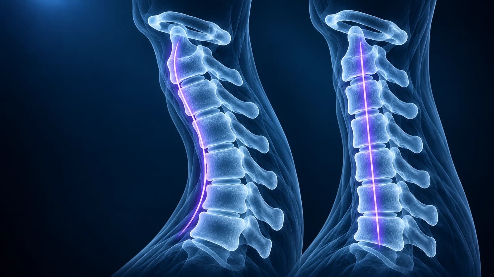
“Straightening of the cervical lordosis” means that on a side view of your neck, the normal gentle curve (hollow at the back of the neck) looks flatter than the textbook shape. It describes posture on that one picture, not a disease. It is common in people with no neck pain at all, and a scan taken lying down, with the chin slightly tucked or with the neck muscles tense, can produce it on its own.
The phrase usually sits in the first lines of a neck X-ray or MRI report, under “alignment”, so many people read it first and worry most about it. This guide explains what radiologists mean by it, the related wording you may see (“loss of lordosis”, “reversal”, “segmental kyphosis”), the situations where the curve genuinely matters, and the findings on the same report that usually deserve more of your attention. It is educational and does not replace the clinician who examined you.
What is the normal cervical lordosis?
Seen from the side, the neck is not a straight column. From the second vertebra (C2) down to the seventh (C7), the bones usually form a soft arc that bulges toward the throat and leaves a hollow at the back of the neck. That arc is the cervical lordosis. It balances the weight of the head over the shoulders and shares load between the discs at the front and the small facet joints at the back.
There is no single “correct” angle. Radiologists and surgeons measure the curve in different ways, for example by drawing lines along the back edges of the vertebral bodies or along the end plates of C2 and C7, and the healthy range between people is wide. Age, sex, the shape of the upper back and simply where the eyes were looking when the image was taken all change the number. Most routine reports therefore describe the curve in words (“maintained”, “straightened”, “reversed”) instead of quoting degrees.
What do the different curve phrases on a neck report mean?
- “Normal cervical lordosis is maintained”: the curve looks typical on this image. Nothing to act on.
- “Straightening” or “loss of the normal cervical lordosis”: the arc is flatter than usual. On its own this is a description, often of position or muscle tension at the moment of imaging.
- “May be positional or related to muscle spasm”: the radiologist is telling you they cannot separate posture from a structural cause on a single image, and is not confirming spasm.
- “Reversal of the cervical lordosis” or “cervical kyphosis”: the curve bends the other way, with the hollow at the front. This is more noteworthy, especially if it is new or sharp.
- “Focal” or “segmental kyphosis at C4–C5”: one disc level is angled forward while the rest of the neck is not. It is often next to a worn disc, but after an injury it can be a sign of a strained or torn ligament at that level.
- “S-shaped” or “sigmoid” alignment: the upper and lower neck curve in opposite directions, a common variant that is usually described without further comment.
For the rest of the wording on the same report, from “uncovertebral hypertrophy” to “T2 hyperintense cord signal”, see our guide to cervical MRI report phrases.
Does a straight neck on X-ray mean muscle spasm?
Not reliably. The old teaching was that painful neck muscles pull the curve straight, and some reports still mention spasm. When researchers in Leeds tested this idea, they found a straight cervical spine in 19% of people arriving at an accident department with acute neck pain and in 26% of people with chronic neck problems, but also in 42% of radiographs from a general population survey, although positioning in that older survey differed. Women were more likely to have a straight neck than men. The authors concluded that their results did not support the idea that a straight neck reflects spasm caused by pain.
In practice, muscle guarding after a strain can flatten the curve for a while, and so can a stiff collar, a pillow under the head or a tucked chin. None of these are visible as such on the image, which is why careful reports use the cautious wording “may be positional”.
Can straightening of the cervical lordosis cause neck pain?
The evidence says the shape alone is a poor explanation for pain. A Swiss study compared side X-rays of 107 volunteers over 45, about half with neck pain in the previous year and half without. The two groups did not differ in overall curvature, in the angle at each level, or in how often the neck was straight or kyphotic. Even a focal kink of more than 4 degrees at one level appeared in 23% of those with pain and 17% of those without, and among people with pain the curve did not track how long, how often or how badly it hurt. The authors advised treating such alignment changes as coincidental unless something else links them to the symptoms.
That matches what clinicians see daily: two people with the same straight neck can feel completely different, and neck pain often settles while the curve on a repeat image looks unchanged. The American College of Radiology notes that age-related changes on neck imaging are common and can mislead in both directions, which is why the history and examination lead, and the image is read in that light.
Why does my MRI show a straight neck when my X-ray did not?
Because the two pictures were taken in different positions. A neck X-ray is usually taken standing or sitting, looking straight ahead, so the head balances on the spine as it does in daily life. An MRI is taken lying on your back with the head resting in a padded coil, often with the chin drawn slightly down to keep still for several minutes. That alone flattens the curve in many people. Comparing curve descriptions between an upright X-ray and a supine MRI, or between two MRIs with different padding, says little about real change. If the curve matters for a decision, clinicians compare like with like, usually upright side X-rays, sometimes with flexion-extension views to see how each level moves.
When does the shape of the neck curve actually matter?
- After an injury: a sharp angle at a single level, widening of the gap between two spinous processes, or one vertebra slipping forward can point to a ligament injury or fracture. Those are judged on CT and sometimes MRI, as described on our pages on whiplash injury and cervical spine fracture.
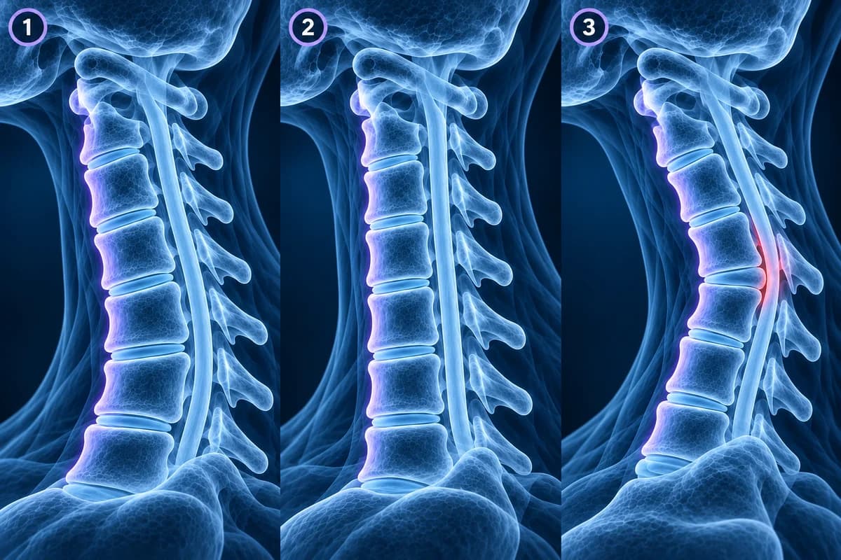
- With spinal cord compression: when discs and bone spurs already narrow the canal, a neck that bends forward drapes the cord over those ridges. A kyphotic neck combined with cord compression is one of the details surgeons weigh in cervical myelopathy, including when choosing a front or back approach.
- After surgery: curve changes above or below a fusion, or a neck that gradually bends forward after the back of several vertebrae was removed, are followed on serial X-rays. Our guide to cervical imaging after fusion covers what is watched.
- With widespread wear: in cervical spondylosis the front of several discs loses height, which can tip the neck toward a flatter or kyphotic shape over years. Here the curve is a consequence of the wear, and the wear findings are what usually matter.
Which findings on the same report usually matter more?
Read past the alignment line. On an MRI, the level-by-level paragraphs say whether a disc osteophyte complex merely touches the fluid around the cord or actually flattens the cord, whether the exit holes for the nerves are narrowed (see cervical foraminal stenosis), and whether the cord shows a bright signal. Those findings, matched with arm symptoms, hand function and walking, guide decisions. A straight curve next to a normal cord and open foramina is a reassuring combination.
Can exercises or a special pillow restore the neck curve?
Programmes promising to “restore the curve” are widely advertised, but because the curve correlates so poorly with pain, a changed angle on a follow-up X-ray is not a useful goal on its own. Treatment for ordinary neck pain aims at symptoms and function: staying active, graded strengthening and mobility work guided by a physiotherapist, attention to long periods of looking down at screens, and pain relief when needed. A pillow that lets you sleep comfortably is worth having for comfort, not to reshape the spine. Ask before starting any manipulation if you have arm symptoms, known cord compression or a recent injury.
When is neck pain with an abnormal curve urgent?
Seek urgent medical care, whatever your report says about the curve, if you have neck pain after a fall, a car crash or a diving injury; weakness, numbness or clumsiness in the arms or hands that is getting worse; a new change in balance or walking; new problems controlling the bladder or bowel; fever with neck pain; a sudden severe headache with a stiff neck; or neck pain with a history of cancer or a weakened immune system.
How can I look at the curve on my own images?
Open the side view of a neck X-ray in our free X-ray viewer, or the middle sagittal slice of an MRI in the free MRI viewer; both run in your browser and keep the files on your device. Follow the back edges of the vertebral bodies from C2 down to C7: in a lordotic neck they trace a smooth arc, in a straightened neck a nearly straight line. Then check the rest of the image rather than stopping there. Our neck X-ray guide walks through the other lines radiologists trace on the same view.
Frequently Asked Questions
Is straightening of the cervical lordosis serious?
On its own, usually not. It is a common description of posture on the image and is often found in people without symptoms. It becomes relevant when it is combined with an injury, cord compression or previous surgery, which your clinician will look at together with your examination.
Is loss of cervical lordosis the same as reversal?
No. Loss or straightening means the curve is flatter than usual. Reversal, or kyphosis, means it bends the opposite way. Reversal attracts more attention, especially if it is sharp, new or at a single level.
Can a straight neck be caused by whiplash?
After a whiplash injury, muscle guarding or a collar can flatten the curve on early images, and it often looks similar in uninjured people too. What matters after trauma is whether any single level is angled, slipped or widened, which points to ligament or bone injury and is assessed with CT or MRI.
Do I need more imaging because my neck curve is straight?
Usually not for that reason alone. Further imaging is driven by symptoms such as worsening arm weakness, hand clumsiness or walking changes, or by a specific question your clinician needs answered.
Can AI explain the alignment section of my neck report?
AI can translate alignment terms and show which part of the image they describe, which helps you prepare questions. It is not a radiologist and cannot judge the curve against your history or examination.
Sources
- Helliwell PS, Evans PF, Wright V. The straight cervical spine: does it indicate muscle spasm? J Bone Joint Surg Br. 1994;76(1):103-106.
- Grob D, Frauenfelder H, Mannion AF. The association between cervical spine curvature and neck pain. Eur Spine J. 2007;16(5):669-678.
- Expert Panel on Neurological Imaging; McDonald MA, Kirsch CFE, et al. ACR Appropriateness Criteria Cervical Neck Pain or Cervical Radiculopathy. J Am Coll Radiol. 2019;16(5S):S57-S76.
Related Articles
What a neck X-ray can and cannot show: the standard views, the four alignment lines, disc space narrowing, bone spurs and the Torg-Pavlov ratio, and when CT or MRI is needed instead.
What your neck MRI report means: uncovertebral hypertrophy, foraminal narrowing grades, effacement of the CSF, cord flattening, T2 hyperintense cord signal, Modic changes and more, each explained in one or two plain sentences.
Why most whiplash MRIs look normal, which injuries MRI can find (ligament tears, bone bruises, disc injury, cord contusion), what report phrases like prevertebral edema mean, and when imaging is needed after a crash.
Related Conditions
Understand your imaging before your next appointment
MRI, X-ray or CT from a CD, folder or ZIP. A PDF report or photos of the films work too.
Free to open and preview, no account needed. An optional AI report from $10 explains your images in plain language, with questions for your doctor.
Your raw files stay on this device. Only rendered images and minimal context are sent when you request an AI report.
Medical Disclaimer: This page is for informational and educational purposes only. It does not constitute medical advice, diagnosis, or treatment. AI-generated analysis may contain errors. Always consult a qualified healthcare professional for medical decisions. Full Disclaimer