
Cervical Flexion-Extension X-ray: What It Shows About Neck Instability
Why flexion and extension neck X-rays are ordered, how the test is done safely, which measurements suggest instability, why they are not the first test after an injury, and what the report wording means.
Key Takeaways
- Flexion-extension X-rays are side views with the head bent forward and back to look for abnormal motion
- More than 3.5 mm of slip or more than 11 degrees of extra angle at one level are widely used warning thresholds
- They add little right after an injury; CT comes first and MRI is used for suspected ligament injury
- You move your own head and stop at once if tingling, weakness or sharp pain starts
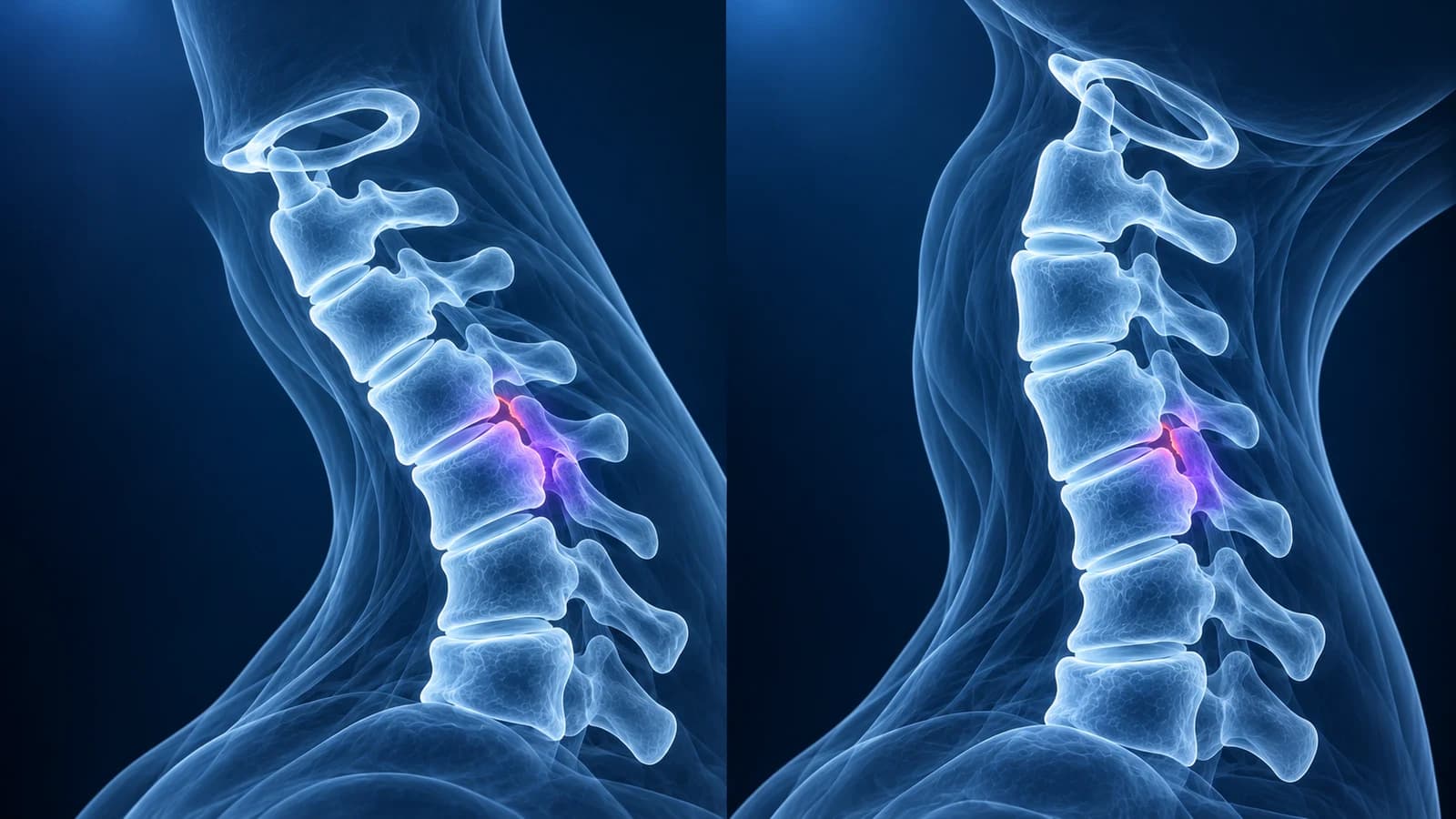
A cervical flexion-extension X-ray is a pair of side views of the neck taken with the head bent forward and then tilted back. It looks for instability: a vertebra that slides or tips more than it should when the neck moves.
Widely used thresholds are more than 3.5 mm of forward or backward slip, or more than 11 degrees of extra angle compared with the neighbouring level. It is a follow-up test for selected questions, not the first scan after an injury, where CT is preferred.
Ordinary neck X-rays and MRI scans are snapshots of a still neck. Some problems, such as a loose ligament or a fusion that has not healed, only show when the neck moves. This guide explains when moving views are useful, how they are taken safely, how to read the report wording and where their limits are. It is educational and not a diagnosis.
Why would a doctor order flexion-extension views of the neck?
- Ongoing neck pain weeks after an injury, once a fracture has been excluded, to check that no ligament was torn enough to let a level move abnormally. Our whiplash injury page covers the wider picture.
- A vertebra already sitting slightly forward on a standing X-ray (“degenerative anterolisthesis”), to see whether it moves further when the neck bends.
- Conditions that loosen the top of the neck, such as rheumatoid arthritis or Down syndrome, where the gap between the first vertebra and the peg of the second is checked in flexion. See atlantoaxial instability.
- After a fusion, to check that the fused level no longer moves and to watch the levels next to it, as described in cervical imaging after fusion.
- Before neck surgery, to plan how many levels need stabilising.
Are flexion-extension X-rays used right after a neck injury?
Rarely. The American College of Radiology prefers CT to radiographs for the first look at a possibly injured neck, and recommends MRI when a ligament injury is suspected. Moving views add little in the first hours or days: pain and protective muscle tension stop the neck from bending far enough to reveal anything, so a “normal” result can be falsely reassuring. In the large NEXUS emergency study, 86 of 818 patients with a confirmed neck injury had flexion-extension views. Only two had a stable bony injury found on those views alone, and four slips seen on them were in patients whose injuries already showed on the standard images. The authors concluded that MRI, CT or delayed moving views fit better when a specific concern remains.
Whether a neck needs any imaging after an injury is decided with validated clinical rules such as the Canadian C-spine rule or the NEXUS criteria, applied by the clinician who examines you. Our pages on cervical spine fracture and what a whiplash MRI shows describe that pathway.
How is a flexion-extension X-ray done safely?
You move your own head; the radiographer never pushes it. You bend forward and back only as far as is comfortable, and you should stop and say so immediately if you feel tingling, electric shocks down the arms, weakness or sharp pain. The test is only done when a clinician has decided that moving the neck is safe, which is why it is not performed on someone who has just been injured and is still being assessed. Each extra side view adds a small radiation dose, similar to the standard neck views.
What measurements suggest cervical instability?
The radiologist compares the two views level by level. Two numbers carry most weight, both tracing back to biomechanical work by White, Panjabi and colleagues in the 1970s:
- Translation: how far the back edge of one vertebral body slides forward or backward relative to the one below. More than 3.5 mm suggests the stabilising ligaments may not be holding.
- Angulation: how much one disc level tips compared with the levels above and below it. More than 11 degrees of difference suggests a loose segment.
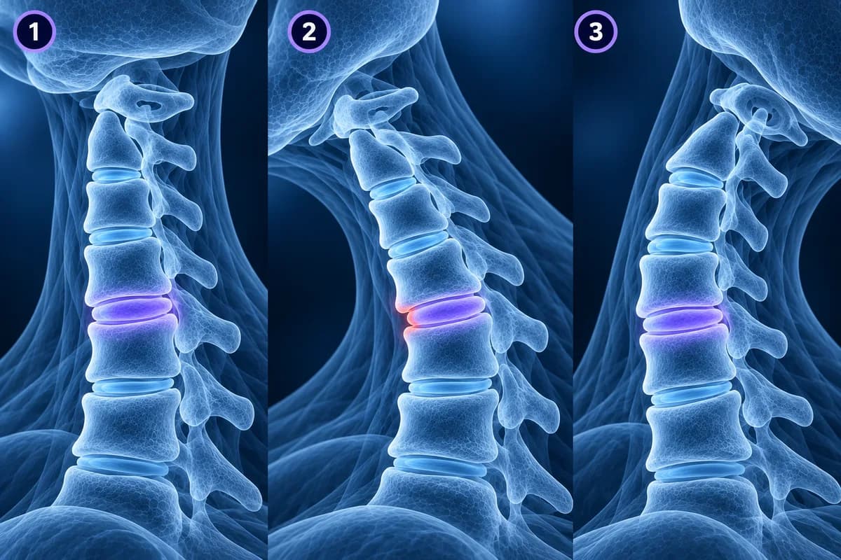
These figures are guides, not pass-or-fail lines. X-ray magnification, how the measurement lines are drawn and how far the neck actually moved all affect them, so borderline values are read together with symptoms, examination and sometimes MRI. At the very top of the neck, a widening gap between the front ring of C1 and the peg of C2 when the head bends forward is the key sign, judged against age-specific limits.
What does the flexion-extension report wording mean?
- “No abnormal motion on flexion and extension”: every level moved within expected limits.
- “Grade 1 anterolisthesis of C4 on C5, 2 mm in neutral, increasing to 4 mm in flexion”: one vertebra sits slightly forward and slides further as the head bends, a dynamic slip worth discussing.
- “Reduces in extension”: the slip returns to normal when the head tilts back, which tells surgeons the level is mobile rather than fixed.
- “Limited range of motion, likely from muscle guarding; instability cannot be excluded”: the neck did not move far enough for the test to answer the question. Repeat views later or an MRI may be suggested.
- “Fanning of the spinous processes” or “widening of the interspinous distance in flexion”: the bony tips at the back of two vertebrae spread apart, a sign of injury to the ligaments joining them.
- “No motion across the fused segment”: after surgery, the fused level is behaving as one block, the expected result.
The neutral view on the same study often carries a comment on the overall curve; our article on straightening of the cervical lordosis explains why that is usually less important than it sounds.
What can flexion-extension X-rays not show?
They show bones only. The spinal cord, the nerves, the discs and the ligaments themselves are invisible, so moving views cannot tell whether a slip is pressing on the cord. For that, MRI is used; a few centres offer upright or dynamic MRI that images the neck in flexion and extension, but it is not widely available. Small fractures are better excluded with CT. The views are also limited by the patient: a stiff, painful or arthritic neck that barely bends gives an incomplete answer. For a full tour of the standard views, see cervical spine X-ray explained.
When should you not wait for a flexion-extension X-ray?
After a significant fall, a road collision or a diving accident, keep the neck still and get emergency care rather than testing its movement. Seek urgent help at any time for numbness, tingling or weakness in the arms or legs, clumsy hands, a change in walking or balance, new bladder or bowel problems, or neck pain with fever, a severe headache, a history of cancer or a weakened immune system.
Can I compare my flexion and extension images myself?
Yes. Open the series in our free X-ray viewer, which keeps the files on your device. Put the flexion and extension views side by side and follow the back edges of the vertebral bodies: in a stable neck they form a smooth line in both positions, while a step that appears or grows in one view is the kind of change the report measures. Our free DICOM viewer also opens CT or MRI from the same visit. Exact millimetre measurements belong to the radiologist, because image scale and line placement change the result.
Frequently Asked Questions
Is a flexion-extension X-ray safe?
It is done only after a clinician has judged that moving the neck is safe, and you control the movement yourself. The radiation dose is small. Tell the radiographer straight away if you feel tingling, weakness or sharp pain.
What does a normal flexion-extension X-ray rule out?
It makes significant instability at the levels shown unlikely, provided the neck moved well. It does not rule out disc problems, nerve or cord compression, or small fractures, and a stiff neck may hide instability.
Why was my flexion-extension X-ray called inconclusive?
Usually because pain or muscle tension limited how far the neck moved, so the levels were not stressed enough to answer the question. Clinicians may repeat the views after a few weeks or choose MRI instead.
Does a slip that increases in flexion mean I need surgery?
Not automatically. Many small degenerative slips are managed without surgery. The decision depends on the amount of movement, whether nerves or the cord are compressed on MRI, and your symptoms and examination.
Can AI measure instability on my neck X-rays?
AI can explain the report wording and point to the levels being compared, which helps you prepare questions. It is not a radiologist, and instability measurements depend on calibrated images and clinical context.
Sources
- White AA 3rd, Johnson RM, Panjabi MM, Southwick WO. Biomechanical analysis of clinical stability in the cervical spine. Clin Orthop Relat Res. 1975;(109):85-96.
- Pollack CV Jr, Hendey GW, Martin DR, et al. Use of flexion-extension radiographs of the cervical spine in blunt trauma. Ann Emerg Med. 2001;38(1):8-11.
- Expert Panel on Neurological Imaging and Musculoskeletal Imaging; Beckmann NM, West OC, et al. ACR Appropriateness Criteria Suspected Spine Trauma. J Am Coll Radiol. 2019;16(5S):S264-S285.
Related Articles
What a neck X-ray can and cannot show: the standard views, the four alignment lines, disc space narrowing, bone spurs and the Torg-Pavlov ratio, and when CT or MRI is needed instead.
Why most whiplash MRIs look normal, which injuries MRI can find (ligament tears, bone bruises, disc injury, cord contusion), what report phrases like prevertebral edema mean, and when imaging is needed after a crash.
What an MRI can and cannot show after an anterior cervical fusion (ACDF): metal artifact, adjacent segment disease, residual compression and cord signal, when CT or X-ray answers better, and which symptoms need urgent care.
Related Conditions
Understand your imaging before your next appointment
MRI, X-ray or CT from a CD, folder or ZIP. A PDF report or photos of the films work too.
Free to open and preview, no account needed. An optional AI report from $10 explains your images in plain language, with questions for your doctor.
Your raw files stay on this device. Only rendered images and minimal context are sent when you request an AI report.
Medical Disclaimer: This page is for informational and educational purposes only. It does not constitute medical advice, diagnosis, or treatment. AI-generated analysis may contain errors. Always consult a qualified healthcare professional for medical decisions. Full Disclaimer