
C5-C6 and C6-C7 Disc Herniation Explained: Symptoms and MRI
Why C5-C6 and C6-C7 are the neck discs that herniate most, which nerve root each one pinches, how the arm symptoms differ, and what protrusion, extrusion and foraminal mean on your MRI.
Key Takeaways
- C5-C6 and C6-C7 are the most common herniation levels in the neck because they move the most
- A C5-C6 disc usually affects the C6 root (thumb side); a C6-C7 disc the C7 root (middle finger)
- Foraminal herniations pinch a root; central ones can compress the spinal cord
- Many cervical herniations improve without surgery; worsening weakness or myelopathy changes that
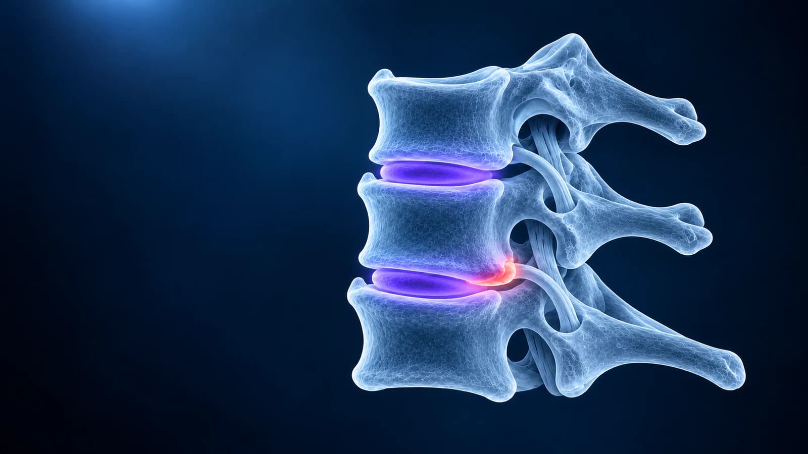
C5-C6 and C6-C7 are the two most common levels for a herniated disc in the neck. A C5-C6 herniation usually irritates the C6 nerve root, causing pain or tingling toward the thumb and index finger and weakness bending the elbow or lifting the wrist. A C6-C7 herniation usually affects the C7 root, sending symptoms toward the middle finger and weakening the triceps. Many of these herniations settle without surgery.
This article explains why these two discs wear first, how to tell the levels apart from your symptoms and your MRI, and what the herniation words in your report mean. The general picture of a slipped disc in the neck is on our cervical disc herniation page. Nothing here is a diagnosis; your clinician’s examination decides which level matters.
Why do C5-C6 and C6-C7 herniate most often?
The lower neck carries the weight of the head and does most of the bending forward and back. The middle and upper levels are stiffer, and the joints at C7-T1 are braced by the ribs and upper back. The two discs in between take the most repeated strain, so they dry out, lose height and crack earliest. In a population study from Rochester, Minnesota, the C7 root was the one most often affected in cervical radiculopathy, followed by C6, which matches these two levels. Herniations at C4-C5 (C5 root) and C7-T1 (C8 root) happen but are less common.
Which nerve root does a C5-C6 or C6-C7 disc pinch?
In the neck each nerve root exits above the vertebra with the same number, so the root is named after the lower of the two bones. A disc that bulges sideways into the nerve exit at C5-C6 meets the C6 root; at C6-C7 it meets the C7 root. A disc that bulges straight back in the middle does not usually pinch a root at all. Instead, it can press on the spinal cord, which produces a different, more serious pattern described below.
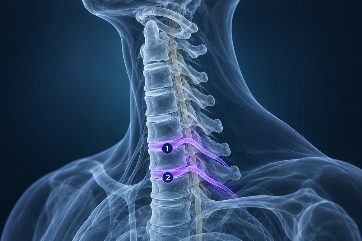
- C6 root (C5-C6 disc): pain from the neck over the shoulder and down the outer forearm, tingling in the thumb and index finger, weaker biceps and wrist extension, reduced biceps or brachioradialis reflex.
- C7 root (C6-C7 disc): pain down the back of the arm, tingling in the middle finger, weaker triceps and finger extension, reduced triceps reflex. Pain between the shoulder blades is common.
- C5 root (C4-C5 disc), for comparison: pain over the shoulder and weakness lifting the arm to the side.
- C8 root (C7-T1 disc), for comparison: tingling in the ring and little fingers and a weaker grip.
These maps overlap between people, so a finding at C5-C6 with symptoms in the middle finger is not necessarily a mismatch. The clinical name for a pinched root with these symptoms is cervical radiculopathy.
What do bulge, protrusion and extrusion mean in a neck MRI report?
Radiologists use a shared vocabulary from the North American Spine Society and radiology societies, written for the lumbar spine and used in the neck too. A “bulge” is a broad, shallow overhang of the disc around more than a quarter of its edge, usually an age change. A herniation is a more focal displacement, and it comes in three shapes:
- Protrusion: the base of the displaced disc is wider than how far it sticks out. Often small.
- Extrusion: the displaced material is wider at its tip than at its base, or has moved up or down behind a vertebra. Extrusions often cause sudden arm pain, and they are also the type most likely to shrink over time.
- Sequestration: a fragment that has broken off completely from the disc, a subtype of extrusion.
The location matters as much as the shape. “Central” points at the cord, “paracentral” is just off the midline and can touch both cord and root, “foraminal” is inside the nerve exit and targets the root, and “extraforaminal” is just outside it. Our cervical MRI report phrases guide covers the rest of the wording.
Is it a soft disc herniation or a disc osteophyte complex?
Younger people more often have a “soft” herniation: fresh disc material that has pushed out, which looks brighter on T2 and can shrink as the body reabsorbs it. In older necks the bulging disc is usually fused with bone spurs from the vertebral edges and the uncovertebral joints, a “hard” ridge that reports call a disc osteophyte complex. It is less likely to shrink, and it is a common cause of narrowed nerve exits (foraminal stenosis) at the same two levels. What that phrase means is covered in disc osteophyte complex explained, and how CT and MRI tell bone from disc is compared in CT vs MRI for cervical radiculopathy.
How do you find a C5-C6 or C6-C7 herniation on your own MRI?
Open the study in the free MRI viewer and pick the sagittal T2 series. Find C2, the tall vertebra with the peg pointing up, and count down: C5-C6 is the fourth disc below C2 and C6-C7 the fifth. A herniation is a dark bump behind the disc pushing into the bright fluid in front of the cord. Then switch to the axial series at the same disc and check which side the bump points to, and whether the nerve exit on that side is darker or smaller than the other. The full method is in how to read a cervical spine MRI.
Can a C5-C6 or C6-C7 herniation compress the spinal cord?
Yes, when it is large and central, or when it adds to a canal that is already narrow. The report then mentions “effacement of the ventral CSF”, “cord flattening” or “cord compression”, and sometimes a bright spot inside the cord. Cord compression causes symptoms in both hands and the legs rather than one arm: clumsy fingers, numb hands, unsteady walking. That pattern is cervical myelopathy, and it changes the treatment discussion.
Does a C5-C6 or C6-C7 herniation need surgery?
Most people with arm pain from a cervical disc herniation are treated without surgery first: time, pain relief, activity changes, physiotherapy and sometimes a steroid injection near the nerve. Many improve over weeks to months. Surgery, most often removing the disc from the front of the neck and fusing the level (ACDF) or replacing the disc, is considered when pain stays disabling despite good non-surgical care, when weakness is significant or progressing, or when the cord is compressed with myelopathy. The decision rests on symptoms and examination, never on the MRI picture alone. Questions about later scans after an operation are covered in cervical MRI after fusion.
When does a neck disc herniation need urgent care?
Get same-day medical care if arm or hand weakness is getting worse, both hands become numb or clumsy, walking or balance changes, bladder or bowel control changes, or the pain started with a significant injury. Fever with severe neck pain, a history of cancer or a weakened immune system also need prompt review, because infection or other causes can mimic a disc. These warning signs matter more than any line in the report.
Frequently Asked Questions
Which is worse, a C5-C6 or a C6-C7 herniation?
Neither level is worse by itself. What matters is the size and position of the herniation, whether it compresses a root or the cord, and how much weakness it causes.
Can I have herniations at both C5-C6 and C6-C7?
Yes, two-level changes are common because the two discs share the same strain. Often only one of them matches the symptoms, and the examination helps identify which.
Can a C6-C7 herniation cause headaches?
Headaches from the neck usually come from the upper levels, C1 to C3. A low-neck herniation more typically causes neck, shoulder-blade and arm pain. See cervicogenic headache.
Will a herniated disc in my neck show on X-ray?
No. An X-ray may show a narrowed disc space or spurs at the level, but the herniation itself needs MRI. See cervical spine X-ray explained.
Can AI tell me which disc is causing my arm pain?
AI can explain where each finding sits and which root that level usually affects. Matching it to your symptoms needs a clinician to test strength, sensation and reflexes.
Sources
- Radhakrishnan K, Litchy WJ, O’Fallon WM, Kurland LT. Epidemiology of cervical radiculopathy. A population-based study from Rochester, Minnesota, 1976 through 1990. Brain. 1994;117(Pt 2):325-335.
- Fardon DF, Williams AL, Dohring EJ, et al. Lumbar disc nomenclature: version 2.0. Recommendations of the combined task forces of the North American Spine Society, the American Society of Spine Radiology and the American Society of Neuroradiology. Spine J. 2014;14(11):2525-2545.
- Expert Panel on Neurological Imaging; McDonald MA, Kirsch CFE, Amin BY, et al. ACR Appropriateness Criteria Cervical Neck Pain or Cervical Radiculopathy. J Am Coll Radiol. 2019;16(5S):S57-S76.
Related Articles
A plain-language guide to your neck MRI: how to count from C2 down to C7, which slices show the cord and the nerve exits, and which artifacts make a cervical scan look worse than it is.
MRI is usually the first scan for a pinched nerve in the neck, while CT shows bone spurs, OPLL and foraminal narrowing in finer detail. Learn when each is used, when CT myelography is needed, and what the report phrases mean.
What an MRI can and cannot show after an anterior cervical fusion (ACDF): metal artifact, adjacent segment disease, residual compression and cord signal, when CT or X-ray answers better, and which symptoms need urgent care.
Understand your imaging before your next appointment
MRI, X-ray or CT from a CD, folder or ZIP. A PDF report or photos of the films work too.
Free to open and preview, no account needed. An optional AI report from $10 explains your images in plain language, with questions for your doctor.
Your raw files stay on this device. Only rendered images and minimal context are sent when you request an AI report.
Medical Disclaimer: This page is for informational and educational purposes only. It does not constitute medical advice, diagnosis, or treatment. AI-generated analysis may contain errors. Always consult a qualified healthcare professional for medical decisions. Full Disclaimer