
Cervical Myelopathy Warning Signs on MRI: Cord Compression and T2 Signal Explained
What cord compression, effaced CSF, T2 hyperintense cord signal and myelomalacia mean on a neck MRI, which symptoms make them urgent, and how doctors weigh MRI findings against clumsy hands and balance problems.
Key Takeaways
- Worsening hand weakness, clumsiness, unsteady walking or bladder and bowel change need same-day medical care
- Cord compression means the cord is deformed with no fluid around it; effaced CSF alone is an earlier stage
- T2 hyperintense cord signal is a bright area inside the cord; a matching dark spot on T1 suggests established damage
- Myelopathy is diagnosed from symptoms and examination, and severity is graded with the mJOA score
- Compression without symptoms is usually followed with education and check-ups rather than preventive surgery
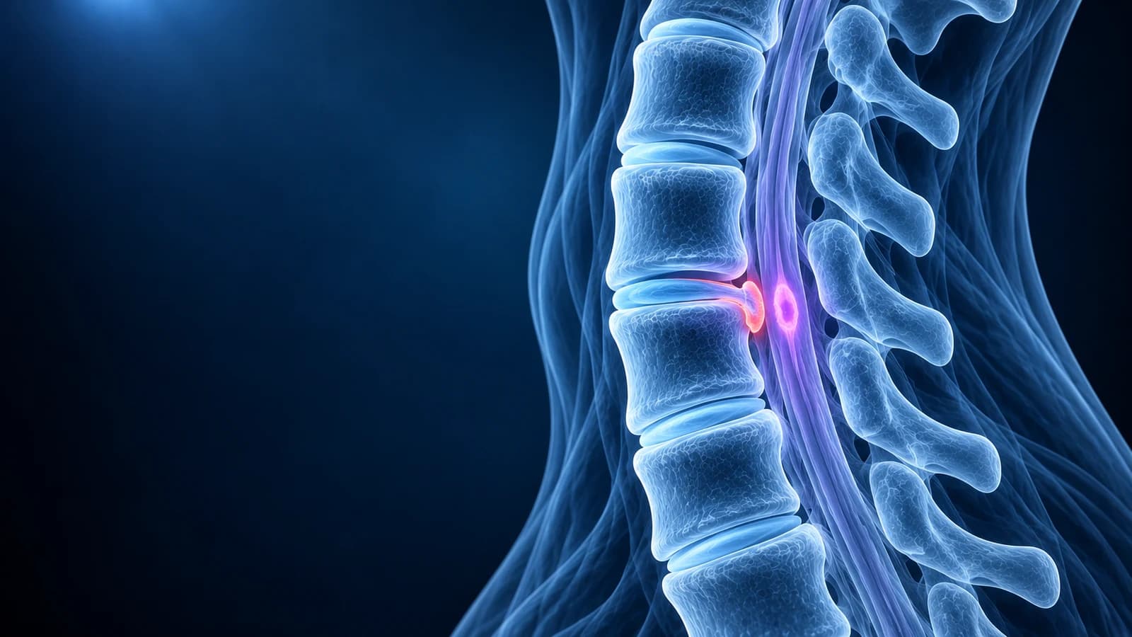
The main MRI warning signs of cervical myelopathy are spinal cord compression, where the cord is flattened with no fluid left around it, and a bright spot inside the cord on T2-weighted images, called “T2 hyperintense cord signal”. A dark spot at the same place on T1 images suggests longer-standing damage. These findings matter most when they match symptoms such as clumsy hands or unsteady walking.
Myelopathy means the spinal cord itself is not working normally. In the neck it is most often caused by slow narrowing of the canal from wear, a combination known as degenerative cervical myelopathy. Unlike a pinched nerve root, which causes pain in one arm, cord compression tends to cause subtle, painless loss of function in both hands and in balance, which is why it is often noticed late. This page explains the MRI phrases that point to it and the symptoms that make them urgent. It is educational and cannot tell you whether you have myelopathy.
Which symptoms mean you should be seen the same day?
Whatever your MRI says, seek urgent medical care, through an emergency department if symptoms are rapid or severe, if you notice any of the following. Do not wait for a scheduled appointment or for a report to be explained.
- Hand or arm weakness that is getting worse over days or weeks
- New clumsiness: dropping objects, trouble with buttons, keys, writing or typing
- Unsteady, stiff or wide-based walking, new falls, or needing to look at your feet to walk
- Numbness in both hands or spreading into the legs, or electric shocks down the spine when bending the neck
- New urgency, hesitancy or loss of control of the bladder or bowel
- Any sudden worsening after a fall or minor neck injury when you already know the canal is narrow
What does cord compression look like on a neck MRI?
In a healthy neck the cord floats in a sleeve of cerebrospinal fluid, which is bright on T2 images and forms a white rim around the grey cord. As discs bulge, spurs grow and the ligament at the back of the canal thickens or buckles, that rim thins and disappears. Radiologists describe the stages in a fairly predictable order, and recognising the stage helps you read how serious a line in the report is.
- “Effacement of the ventral CSF” or “thecal sac indentation” — the fluid in front of the cord is pressed thin, but the cord itself keeps its shape
- “Abuts the cord” or “contacts the ventral cord” — a disc or spur now touches the cord without changing its shape
- “Cord flattening”, “cord deformity” or “cord compression” — the cord is pushed out of its normal oval and no fluid remains around it at that level
- “Severe central canal stenosis” — the canal is narrow enough that the cord is squeezed from front and back, often by a disc osteophyte complex in front and a buckled ligamentum flavum behind
Compression is most common at C5–C6 and C6–C7 and often affects more than one level. Thickened bone along the back of the vertebral bodies, called ossification of the posterior longitudinal ligament (OPLL), can narrow the canal over several levels at once and is best measured on CT.
What does “T2 hyperintense cord signal” mean?
It means a part of the spinal cord looks brighter than the rest on T2-weighted images, usually right at the point of compression. The brightness reflects extra water in the cord tissue: swelling, inflammation, or early loss of tissue from long-standing pressure. Reports may call it “intramedullary signal change”, “cord edema” or “compressive myelopathy changes”.
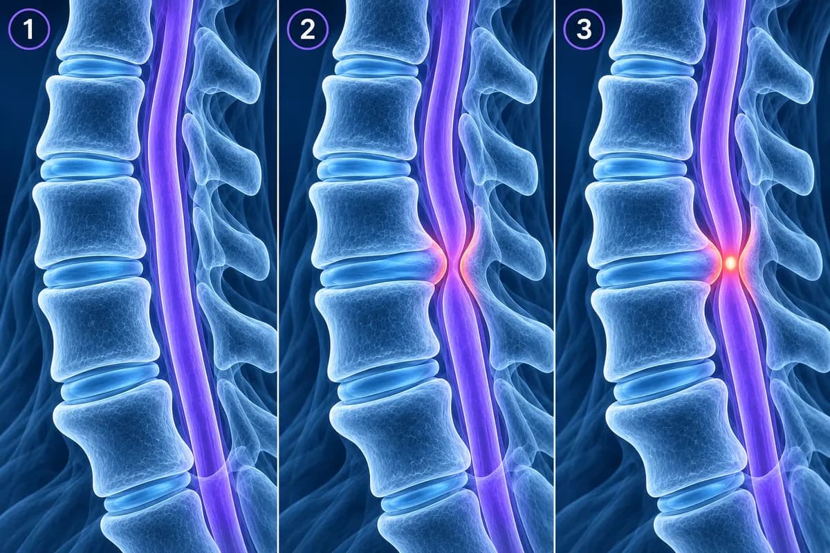
A faint, poorly defined bright area can sometimes improve once pressure is relieved. A sharply outlined, very bright area, especially when the same spot is dark on T1 images, suggests established damage called myelomalacia, which is less likely to recover. On axial images, two small bright dots side by side are sometimes called the “snake-eye” appearance, a pattern associated with longer-standing injury to the grey matter.
Bright cord signal is not specific to compression. Multiple sclerosis, inflammation of the cord, infection, reduced blood supply and some tumours can also cause it, particularly when the bright area is not at a narrowed level. That is why the ACR Appropriateness Criteria for myelopathy treat MRI as the main test and suggest contrast when a non-compressive cause is suspected.
Does cord compression on MRI always mean myelopathy?
No. Myelopathy is a clinical diagnosis: it needs symptoms and examination findings, such as brisk reflexes, a positive Hoffmann sign in the fingers, reduced hand dexterity or a changed gait, that point to the cord. In a large MRI study of 1,211 adults without symptoms, 5.3% had some cord compression and 2.3% had increased cord signal, with both becoming more common after age 50. Compression on a scan with a normal examination is called non-myelopathic cord compression.
The AO Spine clinical practice guideline for degenerative cervical myelopathy treats that situation differently from established myelopathy. For compression without symptoms, it suggests counselling about the risk of progression, teaching the warning signs and clinical follow-up rather than preventive surgery. People who also have arm symptoms from a pinched nerve root are considered at higher risk and should be told so, with surgery or close supervised follow-up both reasonable options.
How do doctors grade cervical myelopathy?
Severity is graded from symptoms, not from the picture. The modified Japanese Orthopaedic Association score, or mJOA, rates upper-limb function, lower-limb function, sensation and bladder function out of 18 points. In the AO Spine guideline, 15 to 17 is mild, 12 to 14 moderate and 11 or below severe. Surgery to decompress the cord is recommended for moderate and severe disease; for mild disease, surgery or a supervised trial of structured rehabilitation are both offered, with surgery advised if function worsens.
MRI informs those decisions by showing how many levels are involved, whether the compression is mainly in front or behind, how the neck is curved, and whether the cord signal suggests reversible or established change. The full clinical picture is described in cervical myelopathy, and the wear that usually drives it in cervical spondylosis.
How can you find the compressed level on your own MRI?
Start with the sagittal T2 series, the side view where the cord runs top to bottom as a grey band inside a white fluid column. Count down from C2, the tall vertebra with the peg pointing up, and look for the level where the white rim disappears or the cord narrows. Then open the axial T2 images at that disc level to see the cord in cross section: a normal cord is a smooth oval, a compressed one looks flattened, kidney-shaped or triangular. Our guide to how to read a cervical spine MRI walks through the sequences step by step.
The free MRI viewer opens your DICOM files in the browser without uploading them, so you can follow along with the report. A layperson looking at slices cannot rule myelopathy in or out, and a bright spot you think you see is not a diagnosis. If you have had surgery, metal causes artefact that changes how the cord looks, as explained in cervical MRI after fusion.
Frequently Asked Questions
Is T2 hyperintense cord signal permanent?
Not always. Faint, poorly defined signal can lessen after the cord is decompressed, while sharply defined signal that is also dark on T1 images is more likely to reflect permanent change. Even then, symptoms often stabilise after treatment. Your spine specialist interprets the signal together with your examination.
Can cervical myelopathy be seen on an X-ray?
An X-ray cannot show the spinal cord, so it cannot confirm myelopathy. It can show features that raise suspicion, such as a congenitally narrow canal, large spurs, OPLL or a slip between vertebrae, and it is often taken alongside MRI to assess alignment and movement before surgery.
What is the difference between myelopathy and radiculopathy?
Radiculopathy is pressure on a single nerve root as it leaves the spine, usually causing pain, tingling or weakness in one arm. Myelopathy is pressure on the spinal cord itself, usually causing clumsy hands, numbness in both hands and balance problems, often with little neck pain. Both can occur together. See cervical radiculopathy for the nerve-root side.
My report says “no cord signal abnormality”. Is that good news?
Yes, it is reassuring. It means the cord tissue looks normal, even if a disc or spur is touching or indenting it. It does not exclude pressure on the cord or nerve roots, so the rest of the report and your symptoms still need to be reviewed with your clinician.
Can a fall make cervical cord compression worse?
Yes. A narrow canal leaves the cord less room, so a fall or a sudden backward bend of the neck can bruise it even without a fracture. Anyone with known compression who develops new weakness or numbness after a fall or minor injury should be assessed urgently.
Sources
- Expert Panel on Neurological Imaging; Agarwal V, Shah LM, Parsons MS, et al. ACR Appropriateness Criteria Myelopathy: 2021 Update. J Am Coll Radiol. 2021;18(5S):S73-S82.
- Fehlings MG, Tetreault LA, Riew KD, et al. A clinical practice guideline for the management of patients with degenerative cervical myelopathy: recommendations for patients with mild, moderate, and severe disease and nonmyelopathic patients with evidence of cord compression. Global Spine J. 2017;7(3 Suppl):70S-83S.
- Davies BM, Mowforth OD, Smith EK, Kotter MR. Degenerative cervical myelopathy. BMJ. 2018;360:k186.
- Nakashima H, Yukawa Y, Suda K, et al. Abnormal findings on magnetic resonance images of the cervical spines in 1211 asymptomatic subjects. Spine (Phila Pa 1976). 2015;40(6):392-398.
Related Articles
A plain-language guide to your neck MRI: how to count from C2 down to C7, which slices show the cord and the nerve exits, and which artifacts make a cervical scan look worse than it is.
A plain-language guide to "disc osteophyte complex" on a cervical MRI: what the ridge is made of, how it differs from a herniated disc, and what effacing the thecal sac, cord contact and cord compression mean.
What an MRI can and cannot show after an anterior cervical fusion (ACDF): metal artifact, adjacent segment disease, residual compression and cord signal, when CT or X-ray answers better, and which symptoms need urgent care.
Related Conditions
Understand your imaging before your next appointment
MRI, X-ray or CT from a CD, folder or ZIP. A PDF report or photos of the films work too.
Free to open and preview, no account needed. An optional AI report from $10 explains your images in plain language, with questions for your doctor.
Your raw files stay on this device. Only rendered images and minimal context are sent when you request an AI report.
Medical Disclaimer: This page is for informational and educational purposes only. It does not constitute medical advice, diagnosis, or treatment. AI-generated analysis may contain errors. Always consult a qualified healthcare professional for medical decisions. Full Disclaimer