
Cervical Foraminal Stenosis: How Neck MRI Grades a Narrowed Nerve Opening
Cervical foraminal stenosis means one of the side openings where a nerve root leaves the neck has become narrow, usually from bone spurs off the uncovertebral and facet joints. Reports grade it mild, moderate or severe. It only matters clinically when the level and side match arm symptoms; narrowing without symptoms is very common after middle age.
Common Symptoms
- Aching or burning pain spreading from the neck over the shoulder blade and into one arm
- Tingling in particular fingers that comes on when looking up, reversing the car or sleeping on one side
- Symptoms that build gradually over months and fluctuate with posture, rather than starting suddenly
- Neck stiffness and reduced rotation toward the affected side
- Mild weakness of grip or of one arm movement, noticed when carrying shopping or working overhead
- Sometimes no symptoms at all, with narrowing found incidentally on a scan done for another reason
When should narrowing in the neck be checked urgently?
Foraminal stenosis affects a nerve root, not the spinal cord, and is rarely an emergency on its own. Get same-day care if arm weakness is worsening, if both hands become clumsy or numb, if walking or balance changes, or if you notice new bladder or bowel problems. Those features suggest the central canal and cord may be involved, which is covered on our cervical myelopathy page. Neck pain after significant trauma, with fever, or with a history of cancer also needs prompt assessment.
What is the neural foramen in the neck, and why does it narrow?
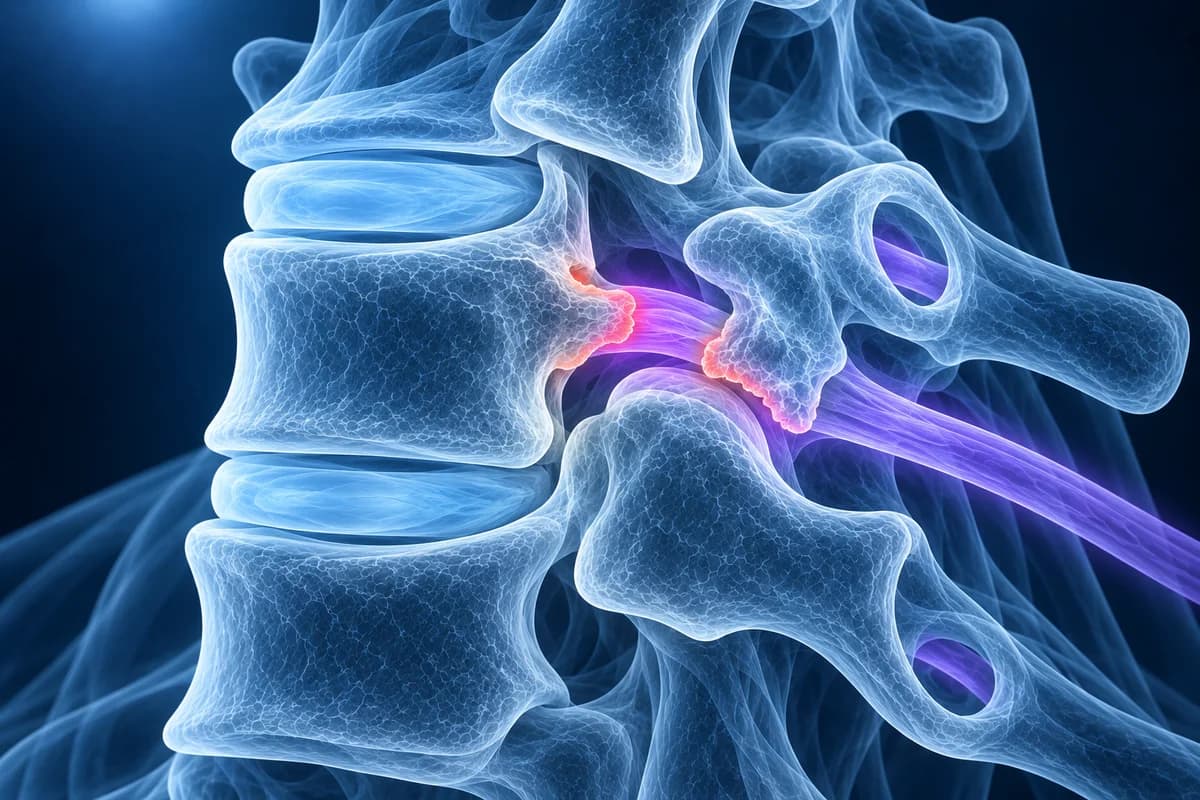
Between each pair of cervical vertebrae there is an opening on the left and one on the right, called the neural foramen (plural foramina). The nerve root for that level passes through it on its way to the shoulder, arm and hand. In the neck these openings are short tunnels angled forward and outward at roughly 45 degrees, which is why a straight side view does not show them well.
Each opening has bony walls on all sides. The front wall is formed by the uncovertebral joint, a small joint unique to the cervical spine that sits at the back corner of each vertebral body from C3 to C7. The back wall is the facet joint. As the disc between the vertebrae loses height with age, the vertebrae settle closer together, the uncovertebral joints rub and grow spurs, the facet joints enlarge, and the opening shrinks from several directions at once. A bulging disc can add to the narrowing from the front. This combination is part of the broader picture of cervical spondylosis.
The C5–C6 and C6–C7 openings are the ones most often narrowed, because those levels move the most. When narrowing squeezes the root enough to cause pain, tingling or weakness down the arm, the clinical diagnosis becomes cervical radiculopathy.
What do mild, moderate and severe foraminal stenosis mean?
There is no single universal scale, so radiologists usually describe how much of the fat around the nerve remains and how close bone or disc comes to the root. A widely cited MRI grading system on axial T2 images (Kim and colleagues, 2015) compares the narrowest width of the opening with the width of the nerve root just outside it:
- Grade 0, no stenosis: the narrowest part of the opening is wider than the nerve root.
- Grade 1, non-severe stenosis: the opening is the same width as the root or narrower, but still more than half its width.
- Grade 2, severe stenosis: the opening is half the width of the root or less.
Another scheme uses oblique sagittal MRI images, cut along the tunnel, to grade from 0 to 3. In everyday reports these usually translate into "mild" (some fat still surrounds the root), "moderate" (fat mostly gone, root contacted) and "severe" (root flattened or its outline lost). Grading varies between readers, so two reports of the same scan can differ by one step.
Which report phrases describe cervical foraminal stenosis?
- "Uncovertebral hypertrophy" or "uncinate spurring"
- Bony overgrowth of the uncovertebral joint at the front of the nerve opening. It is the single most common cause of cervical foraminal narrowing.
- "Facet arthropathy" or "facet hypertrophy"
- Wear and enlargement of the facet joint that forms the back wall of the opening. Combined with uncovertebral spurs, it pinches the foramen from front and back.
- "Bilateral foraminal narrowing"
- Both the left and right openings at that level are narrowed. Symptoms are often on one side only even when the scan shows both.
- "Effacement of the perineural fat"
- The fat cushion that normally surrounds the root in the opening is no longer visible. It is the main sign radiologists use to call narrowing moderate or severe.
- "Disc osteophyte complex extending into the foramen"
- A ridge of disc and bone spur at the back of the disc that reaches sideways into the opening. Read more in disc osteophyte complex explained.
For the rest of a typical neck report, see cervical MRI report phrases explained.
Why are oblique X-rays or CT used for foraminal stenosis?
Because the cervical foramina point forward and outward, a standard side-on X-ray looks across them at an angle. Oblique X-ray views are tilted to line up with the tunnels, so the opening appears as an oval and spurs bulging into it are easy to see. Our guide to the cervical spine X-ray explains these views.
CT shows bone edges more sharply than any other test and reconstructions can be angled through each foramen, so it is often used to separate a hard bony spur from a soft disc before surgery. MRI shows the nerve root and disc and is the usual first advanced test; oblique sagittal MRI slices improve its view of the foramina. Some MRI sequences, especially gradient-echo images, can make bone spurs look larger than they are and overstate the narrowing. The ACR Appropriateness Criteria for cervical radiculopathy list MRI without contrast and X-rays among the first studies, with CT or CT myelography when MRI cannot be done or leaves the question open. See CT vs MRI for cervical radiculopathy for the trade-offs.
What we look for on foraminal stenosis imaging
- The grade of narrowing at each level and side, from C2–C3 down to C7–T1
- Whether uncovertebral spurs, facet overgrowth, disc bulge or a combination is causing it
- Loss of perineural fat and flattening of the exiting nerve root on axial and oblique images
- Loss of disc height that lets the vertebrae settle and the openings shrink
- Associated central canal narrowing or contact with the spinal cord
- Slight slippage of one vertebra on another, which can narrow an opening further with movement
Is severe foraminal stenosis on my MRI dangerous?
Not by itself. A severely narrowed opening can be completely painless, and a mild one can cause symptoms if the root is inflamed. Clinicians use the grade together with your symptoms, strength and reflexes. Severe narrowing that matches arm pain and weakness is more likely to respond to a procedure that widens the opening, while severe narrowing on the side without symptoms is usually just watched.
If the report also mentions the central canal or the spinal cord, read that line carefully, because cord compression is managed differently from root compression. Calcified or ossified ligaments behind the vertebral bodies, as in OPLL, can narrow both the canal and the foramina.
How is cervical foraminal stenosis treated?
Treatment follows symptoms, not the scan grade. Physiotherapy focused on posture, neck and shoulder blade strength, and avoiding long periods of looking up often helps. Pain relief and, for persistent arm pain, an image guided nerve root injection are options a clinician may offer. Because bony narrowing does not shrink on its own the way a soft disc can, symptoms tend to fluctuate rather than vanish.
When pain or weakness persists, surgery can widen the opening. A posterior cervical foraminotomy removes part of the facet joint from behind to free the root and keeps motion at that level. Anterior cervical discectomy and fusion (ACDF) removes the disc and spurs from the front and restores disc height, which also opens the foramen; cervical disc replacement is an alternative for some patients. Your spine surgeon will match the option to the cause and location of the narrowing. Narrowing of the openings in the lower back produces leg symptoms and is a separate lumbar topic.
How can I see my own nerve openings on a neck scan?
Open your MRI in the free cervical MRI viewer and scroll the axial slices at the reported level, comparing left and right openings. X-rays, including oblique views, open in the free X-ray viewer, and CT in the free CT viewer. Files stay on your device. For a plain-language explanation of the grades in your report, upload your imaging for an AI review. It explains findings; it is not a diagnosis and does not replace your radiologist or spine specialist.
Frequently Asked Questions
What does moderate foraminal stenosis at C5–C6 mean?
The opening where the C6 nerve root leaves the spine at C5–C6 is clearly narrowed, with most of the surrounding fat gone, but the root is not severely flattened. It may or may not cause symptoms; it is relevant if you have pain or tingling toward the thumb side of the arm on the same side.
Can foraminal stenosis get better without surgery?
The bony narrowing itself does not reverse, but symptoms often settle as nerve inflammation calms down, with physiotherapy, activity changes and pain management. Many people live with narrowing on their scans and no ongoing symptoms.
Is foraminal stenosis the same as spinal stenosis?
No. Spinal or central stenosis is narrowing of the main canal that holds the spinal cord. Foraminal stenosis is narrowing of the side opening for a single nerve root. They can occur together at the same level, and central narrowing in the neck carries the risk of cord compression.
Why do two radiologists grade my foraminal stenosis differently?
Grading depends on which slices and sequences are used and on reader judgement, and in the 2015 axial MRI grading study, agreement between different readers was only fair to moderate. A one-step difference, such as mild versus moderate, is common and rarely changes management on its own.
Should I avoid exercise if I have foraminal stenosis?
Most people are encouraged to stay active. Movements that close the opening, such as prolonged looking up or tilting toward the painful side, may need adjusting. Ask your clinician or physiotherapist to tailor exercises, especially if you have weakness or any signs of cord involvement.
Sources
- Kim S, Lee JW, Chai JW, et al. A New MRI Grading System for Cervical Foraminal Stenosis Based on Axial T2-Weighted Images. Korean J Radiol. 2015;16(6):1294-1302.
- Park HJ, Kim SS, Lee SY, et al. A practical MRI grading system for cervical foraminal stenosis based on oblique sagittal images. Br J Radiol. 2013;86(1025):20120515.
- Expert Panel on Neurological Imaging; McDonald MA, Kirsch CFE, Amin BY, et al. ACR Appropriateness Criteria Cervical Neck Pain or Cervical Radiculopathy. J Am Coll Radiol. 2019;16(5S):S57-S76.
- Margetis K, Magnus W, Mesfin FB. Cervical Radiculopathy. StatPearls. Treasure Island (FL): StatPearls Publishing.
Related Articles
Why C5-C6 and C6-C7 are the neck discs that herniate most, which nerve root each one pinches, how the arm symptoms differ, and what protrusion, extrusion and foraminal mean on your MRI.
MRI is usually the first scan for a pinched nerve in the neck, while CT shows bone spurs, OPLL and foraminal narrowing in finer detail. Learn when each is used, when CT myelography is needed, and what the report phrases mean.
What your neck MRI report means: uncovertebral hypertrophy, foraminal narrowing grades, effacement of the CSF, cord flattening, T2 hyperintense cord signal, Modic changes and more, each explained in one or two plain sentences.
Understand your imaging before your next appointment
MRI, X-ray or CT from a CD, folder or ZIP. A PDF report or photos of the films work too.
Free to open and preview, no account needed. An optional AI report from $10 explains your images in plain language, with questions for your doctor.
Your raw files stay on this device. Only rendered images and minimal context are sent when you request an AI report.
Medical Disclaimer: This page is for informational and educational purposes only. It does not constitute medical advice, diagnosis, or treatment. AI-generated analysis may contain errors. Always consult a qualified healthcare professional for medical decisions. Full Disclaimer