
Cervical Spine Fracture: Emergency Signs, CT Findings and Stability
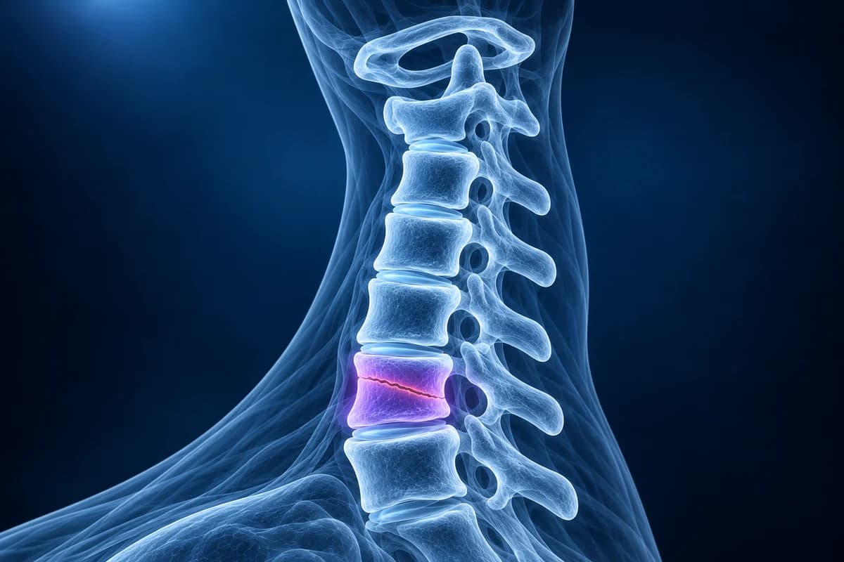
A cervical spine fracture is a break in one of the seven neck vertebrae, C1 to C7. It is a medical emergency until a clinician proves otherwise, because an unstable fracture can injure the spinal cord. In adults who need imaging after neck trauma, CT is the first test; MRI is added to check the ligaments, discs and cord. Many fractures are stable and heal in a collar, while unstable ones need surgery.
Some signs described on this page need prompt medical care. If you have them, get care first. Reading about imaging can wait.
Common Symptoms
- Severe pain in the middle of the back of the neck, directly over the bones, after a fall, crash or dive
- Unwillingness or inability to turn the head, often holding the head with the hands
- Numbness, tingling, burning or electric shocks in the arms, hands or legs
- Weakness of the arms or legs, clumsy hands, or difficulty standing and walking
- Loss of bladder or bowel control, or reduced sensation around the groin
- In older adults, new neck pain after a simple fall from standing height, sometimes with little else
Could my neck be broken? Get urgent care now if:
- You have neck pain after a car crash, fall from a height or down stairs, diving into shallow water, a blow to the head, or a sports collision.
- You feel numbness, tingling, weakness or electric shocks in any limb, or your hands suddenly feel clumsy.
- You have trouble walking, lose your balance, or notice a change in bladder or bowel control.
- You are 65 or older and hurt your neck in any fall, or you have ankylosing spondylitis, DISH or a previous neck fusion, even if the injury seemed minor.
- You have a severe headache, double vision, slurred speech or difficulty swallowing after neck trauma, which can signal injury to an artery in the neck.
Call emergency services rather than driving yourself. Keep the head and neck still, do not let anyone pull or manipulate the neck, and do not remove a helmet unless breathing is at risk. An online scan review is never a substitute for an emergency assessment.
Who needs a scan after a neck injury?
Two validated decision rules help emergency clinicians decide who needs imaging. Under the NEXUS criteria, a patient can be cleared without imaging only if all five are true: no tenderness over the midline of the back of the neck, no focal neurological deficit, normal alertness, no intoxication, and no painful distracting injury elsewhere. The Canadian C-spine rule, for alert and stable adults, adds a check of age (65 or over), a dangerous mechanism and limb tingling, then asks low-risk patients to actively rotate the neck 45 degrees each way.
These rules are applied by a clinician who has examined you. They are not self-tests, and they do not apply to people who are drowsy, intoxicated, distracted by another painful injury, or to young children, who are assessed with separate pathways.
Why is CT the first test for a suspected neck fracture?
The ACR Appropriateness Criteria for suspected spine trauma rate non-contrast CT of the cervical spine as the imaging test of choice for adults who meet criteria for imaging. CT is fast, shows fine bone detail in every plane, and is far better than plain X-rays at finding fractures at the skull base, C1 and C2, and the junction with the chest at C7 and T1, where shoulders obscure radiographs. The axial slices are reconstructed into sagittal and coronal views so alignment can be traced along the front and back of the vertebral bodies and through the facet joints.
MRI is added when there are neurological symptoms, when CT is normal but pain or examination still suggest a ligament injury, before surgery, and in people with a rigid, fused spine. CT angiography of the neck vessels is considered when a fracture runs through the transverse foramen that carries the vertebral artery, or when facets are dislocated. Plain X-rays still have a role later, for follow-up of healing and, once a fracture is excluded, for movement views such as those in our flexion-extension X-ray guide. The cervical spine X-ray explainer shows the alignment lines radiologists trace.
What are the main types of neck fracture?
The upper neck (C1 and C2) and the lower neck (C3 to C7) break in different ways. In the upper neck, a Jefferson fracture is a burst of the C1 ring from a blow to the top of the head; its stability depends on whether the transverse ligament that holds the dens is torn, which is the same structure discussed on our atlantoaxial instability page. Odontoid fractures break the upward peg of C2 and are graded type I (tip), type II (base of the peg, the most common and the most prone to healing problems) and type III (extending into the body of C2). A hangman's fracture breaks both sides of the C2 arch, typically from forced extension.
In the lower neck, compression and burst fractures flatten the vertebral body, flexion teardrop fractures combine a front corner fragment with ligament disruption and are highly unstable, and facet dislocations lock one or both facets forward. Some fractures are minor: an isolated spinous process fracture at C7, the clay-shoveler's fracture, or a transverse process fracture away from the artery is usually stable. Surgeons often summarise lower-neck injuries with the Subaxial Injury Classification, which combines the fracture shape, the integrity of the ligaments and the neurological status.
What does my cervical CT report mean?
- "Nondisplaced fracture" versus "displaced fracture"
- Nondisplaced means the bone is cracked but the pieces are still lined up. Displaced means the pieces have moved apart, which raises concern for instability and nerve or cord injury.
- "Retropulsion" or "retropulsed fragment"
- A piece of the vertebral body has been pushed backward into the spinal canal, toward the cord. The report usually states how much of the canal is narrowed.
- "Perched facet" or "jumped facet"
- The small joints at the back of the spine have slid forward so that the upper facet sits on top of (perched) or in front of (jumped, locked) the lower one. This is a dislocation and usually needs urgent reduction.
- "Fracture extends into the transverse foramen"
- The break involves the small bony tunnel that carries the vertebral artery, so the artery is often checked with CT angiography.
- "Prevertebral soft tissue swelling"
- Thickening of the tissue in front of the vertebrae from bleeding or edema. It can be the only clue to a ligament injury or a subtle fracture.
- "Chronic-appearing" or "corticated fragment"
- A smooth, rounded bone fragment with its own outer rim, which suggests an old injury or a normal variant rather than a fresh fracture.
For MRI wording such as cord edema or ligament disruption, see our cervical MRI report phrases guide and how to read a cervical spine MRI.
What we look for on neck trauma imaging
- Cortical breaks in the vertebral body, arch, pedicles, lateral masses, dens or spinous processes
- Step-offs in the anterior and posterior vertebral lines and the spinolaminar line
- Facet joint widening, perched or locked facets, and loss of facet joint congruity
- Widening of the gap between C1 and the dens or between adjacent spinous processes
- Retropulsed fragments narrowing the canal and fractures crossing the transverse foramen
- On MRI, ligament disruption, traumatic disc herniation and T2 hyperintense cord signal
Why are older adults and people with a stiff spine at higher risk?
In older adults with osteoporosis, a fall from standing height is enough to break the upper neck, and type II odontoid fractures are a classic result. Symptoms can be surprisingly mild, which is why age alone triggers imaging under the Canadian C-spine rule. Background wear such as cervical spondylosis can also hide subtle fractures among osteophytes on X-ray, another reason CT is preferred.
A spine fused by ankylosing spondylitis or bridged by diffuse idiopathic skeletal hyperostosis (DISH) behaves like a long bone: it can snap straight across after a minor fall, and these fractures are often unstable and easy to miss. Many centres image the whole spine with CT and MRI in these patients after any trauma. People with a narrow canal from degeneration can also suffer cord injury from an extension injury without any fracture; the cord findings are described on our cervical myelopathy page.
How are cervical fractures treated?
Treatment depends on stability, alignment and the neurological examination. Stable fractures without nerve involvement are usually treated in a rigid collar for several weeks, with follow-up X-rays or CT to confirm healing. Some upper-neck injuries are held in a halo frame. Unstable fractures, dislocations and fractures with cord compression are usually treated surgically by realigning and fixing the spine with screws, rods or plates, and when the cord is compressed, by decompressing it. Patients with a spinal cord injury are managed in specialist units; that topic is covered in depth on the general spine site, not here.
Can I look at my own neck CT after the emergency visit?
Once you have been assessed and treated, you can open your images privately in the free neck CT viewer, or the free X-ray viewer for follow-up films. Files stay on your device. To understand the wording of your report, upload the images for an AI explanation. It is not a diagnosis, it cannot clear your neck, and it never replaces your emergency team or spine surgeon.
Frequently Asked Questions
Can you walk around with a broken neck?
Yes. Many people with a stable cervical fracture walk into the emergency department, and some unstable fractures also produce no nerve symptoms at first. Being able to walk or move the arms does not rule out a fracture, which is why neck pain after significant trauma needs a clinical assessment and, when the decision rules say so, a CT.
Can a neck X-ray miss a fracture?
Yes. Plain X-rays can miss fractures, especially at C1 and C2 and where the shoulders overlap C7 and T1. That is why the ACR recommends CT rather than X-rays as the first test for adults who need imaging after neck trauma.
Why did I need an MRI after a normal CT?
CT shows bone very well but shows ligaments, discs and the spinal cord poorly. MRI is added when there are nerve symptoms, when pain and tenderness persist despite a normal CT, or when the spine is fused by disease. It looks for ligament tears, disc injury, bleeding in the canal and bruising or swelling of the cord.
How long does a neck fracture take to heal?
Bone healing usually takes around two to three months, during which a collar or brace is often worn. Odontoid fractures in older adults may heal with fibrous tissue instead of bone, which can still be stable. After surgery the timeline depends on the fixation. Your spine team decides when to remove the collar, usually after follow-up imaging.
What does "no acute fracture or malalignment" mean on a neck CT?
It means the radiologist saw no fresh break and the vertebrae are lined up normally. It does not exclude a ligament injury, which CT cannot see directly, so ongoing pain or any nerve symptoms should still be reviewed.
Sources
- Expert Panel on Neurological Imaging and Musculoskeletal Imaging; Beckmann NM, West OC, et al. ACR Appropriateness Criteria Suspected Spine Trauma. J Am Coll Radiol. 2019;16(5S):S264-S285.
- Hoffman JR, Mower WR, Wolfson AB, et al. Validity of a set of clinical criteria to rule out injury to the cervical spine in patients with blunt trauma. N Engl J Med. 2000;343(2):94-99.
- Stiell IG, Wells GA, Vandemheen KL, et al. The Canadian C-spine rule for radiography in alert and stable trauma patients. JAMA. 2001;286(15):1841-1848.
Related Articles
Why most whiplash MRIs look normal, which injuries MRI can find (ligament tears, bone bruises, disc injury, cord contusion), what report phrases like prevertebral edema mean, and when imaging is needed after a crash.
Why flexion and extension neck X-rays are ordered, how the test is done safely, which measurements suggest instability, why they are not the first test after an injury, and what the report wording means.
What a neck X-ray can and cannot show: the standard views, the four alignment lines, disc space narrowing, bone spurs and the Torg-Pavlov ratio, and when CT or MRI is needed instead.
Do not delay urgent medical care to use this tool.
After you get care, understand your imaging
MRI, X-ray or CT from a CD, folder or ZIP. A PDF report or photos of the films work too.
Free to open and preview, no account needed. An optional AI report from $10 explains your images in plain language, with questions for your doctor.
Your raw files stay on this device. Only rendered images and minimal context are sent when you request an AI report.
Medical Disclaimer: This page is for informational and educational purposes only. It does not constitute medical advice, diagnosis, or treatment. AI-generated analysis may contain errors. Always consult a qualified healthcare professional for medical decisions. Full Disclaimer