
How to Read a Cervical Spine MRI: Levels, Planes and Pitfalls
A plain-language guide to your neck MRI: how to count from C2 down to C7, which slices show the cord and the nerve exits, and which artifacts make a cervical scan look worse than it is.
Key Takeaways
- Count levels from C2, the vertebra with the upward-pointing dens, down to C7-T1
- In the neck, the C6 root exits at C5-C6 and the C7 root at C6-C7
- Check every disc for fluid around the cord, open nerve exits and normal cord signal on T2
- Gradient echo images can exaggerate bone spurs, and bulges are common in people without symptoms
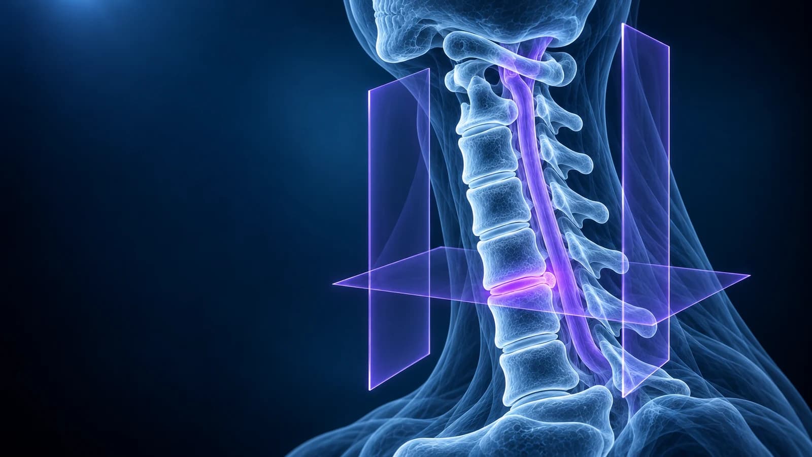
To read a cervical spine MRI, first label the levels by counting down from C2, then check three things at every disc from C2-C3 to C7-T1: is there still a rim of bright fluid around the spinal cord, are the two nerve exits (foramina) open, and does the cord itself look uniformly grey on T2 images? Most neck MRI reports are organised exactly this way, one disc level per line.
The neck differs from the lower back in ways that change how a scan is read. The canal contains the spinal cord rather than a bundle of loose nerve roots, the nerve exits point forward at an angle, and small joints called uncovertebral joints line the sides of each disc. This guide covers only the cervical-specific parts; general MRI physics and lumbar findings are covered on the whole-spine guides of our sister spine site. It is educational and does not replace the radiologist’s report or your clinician’s exam.
How do you count the levels on a neck MRI?
Counting is easier in the neck than anywhere else in the spine because the top is unmistakable. C1, the atlas, is a thin ring with no real body. C2, the axis, is the tall vertebra whose peg (the dens or odontoid process) points up through that ring. Everything below is counted from C2: C3, C4, C5, C6 and C7, then the first thoracic vertebra. Disc levels are named after the two bones they sit between, so “C5-C6” is the disc under C5 and above C6.
The nerve numbering is the part that trips people up. In the neck, each nerve root leaves above the vertebra that shares its number, so the C6 root exits at C5-C6 and the C7 root at C6-C7. There are eight cervical nerve roots but only seven cervical vertebrae, which is why the C8 root exits at C7-T1. A report that mentions “left C5-C6 foraminal narrowing” is therefore talking about the left C6 nerve root. The two levels that cause most arm symptoms are compared side by side in C5-C6 and C6-C7 disc herniation explained.
Which sequences and planes are in a cervical MRI?
- Sagittal T2: the main overview. Spinal fluid is bright, the cord is mid-grey, discs are grey to dark when they dry out. This is where cord flattening and bright cord signal are judged.
- Sagittal T1: anatomy and bone marrow. Healthy marrow is bright; a dark area replacing it suggests edema, infection, fracture or another marrow process that the report should name.
- Sagittal STIR: fat is switched off, so marrow edema, ligament injury after trauma and swelling around the discs stand out.
- Axial T2 or gradient echo (often labelled T2* or MEDIC): slices straight across each disc. Gradient echo makes the disc and the cord easy to separate from bone, which is why many centres use it in the neck.
- Oblique sagittal (only at some centres): slices angled along the nerve exits, which run forward and outward at roughly 45 degrees, so each foramen is seen as a keyhole along its length.
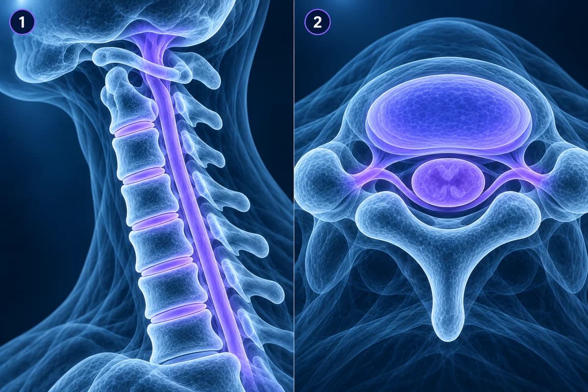
Contrast (gadolinium) is not part of a routine neck MRI. It is added when infection such as discitis-osteomyelitis, a tumour, inflammation of the cord or previous surgery is the question.
What should the spinal cord look like on a neck MRI?
On sagittal T2 the cord is a smooth grey ribbon floating in a sleeve of bright fluid, slightly wider around C5 and C6 where the nerves to the arms leave. Radiologists check two things. First, space: whether the bright fluid in front of and behind the cord is preserved, “effaced” (pushed away), or gone with the cord pressed flat. Second, signal: whether a bright patch appears inside the cord itself, reported as “T2 hyperintense cord signal” or “myelomalacia” when it looks old.
Cord compression with symptoms such as clumsy hands or unsteady walking is called cervical myelopathy, and it is the finding on a neck MRI that most needs prompt follow-up. A grading system published in 2011 sorts canal narrowing into four steps, from fluid partly lost (grade 1), to the cord deformed (grade 2), to bright cord signal (grade 3). What each step looks like is shown in cervical myelopathy warning signs on MRI.
How do you look at the nerve exits (foramina) on axial slices?
On an axial slice through a cervical disc, the canal sits in the middle like a triangle and the two foramina angle forward and outward from its front corners. Each foramen is bordered in front by the uncovertebral joint and the disc edge, and behind by the facet joint. That is why the two most common causes of a narrowed exit in the neck are bone spurs from these small joints (“uncovertebral hypertrophy” and “facet hypertrophy”) and a disc that bulges sideways, rather than the large soft herniations typical lower down.
Because the foramen runs at an angle, a single axial slice cuts across it obliquely and can make it look narrower than it is. Scroll one or two slices above and below the disc and compare right with left. A tight exit on the same side as your arm symptoms fits cervical foraminal stenosis, and a pinched root producing pain, tingling or weakness down the arm is cervical radiculopathy. Foraminal narrowing on the opposite side to your symptoms is usually an incidental finding.
Which artifacts make a cervical MRI look worse than it is?
- Gradient echo blooming: on T2* axial images, bone spurs look larger than they really are, so foraminal narrowing can be overestimated. Radiologists compare with the other sequences before grading.
- Truncation (Gibbs) artifact: a thin bright or dark line running down the middle of the cord on sagittal images can mimic a fluid-filled cavity (syrinx). It usually disappears on axial slices.
- Swallowing and breathing motion: blurring in front of the spine and ghost copies of the throat can smear across the canal at a single level.
- CSF flow: pulsing spinal fluid can create dark swirls behind the cord that look like a mass but change shape from slice to slice.
- Neck position: the scan is taken lying flat with the neck neutral, so narrowing that only appears when bending or extending may be under-called. Movement-related questions are what flexion-extension X-rays are for.
Are disc bulges on a neck MRI normal?
Very often, yes. A Japanese study of 1,211 people with no neck symptoms found disc bulging in most of them, more frequent with age and most often at C5-C6, and a small minority had some cord compression without any symptoms. So a report listing a bulge at every level describes cervical spondylosis that is common with age, not necessarily the source of your pain. What matters is whether a specific level matches your symptoms and your examination. Phrases such as “loss of the normal cervical lordosis” are similarly common and explained in what straightening of the cervical lordosis means.
When should neck MRI findings be checked urgently?
Seek same-day medical care, whatever the report says, if you notice arm or hand weakness that is getting worse, clumsy hands (dropping things, trouble with buttons), a change in walking or balance, new bladder or bowel problems, fever with neck pain, or neck pain after a significant fall or crash. A history of cancer or a weakened immune system also lowers the threshold for urgent review. Reading your own scan is never a reason to delay that care.
Can I open my own cervical MRI images?
Yes. Load the DICOM folder from your disc into the free MRI viewer, which opens it in your browser without uploading the original files. Choose the sagittal T2 series, find C2, count down to the level your report names, then switch to the axial series at the same disc. Keep the report beside you; our cervical MRI report phrases guide translates each line.
Frequently Asked Questions
Why does my neck MRI report list every disc level separately?
Because each disc can look different, and each level corresponds to a different nerve root. Radiologists describe the disc, the central canal and both foramina at every level from C2-C3 to C7-T1, then summarise the most important finding in the impression at the end.
Does a cervical MRI show the upper back too?
Usually only the top two or three thoracic vertebrae appear at the bottom of the sagittal slices, and they may not be reported in detail. Problems lower in the upper back need a thoracic MRI.
Can a neck MRI miss a fracture?
MRI shows ligament injury and bone edema well, but CT shows fine fracture lines better. After significant trauma, CT is the usual first test in adults who need imaging, and MRI is added when the cord or ligaments need assessing. See cervical spine fracture.
Do I need an MRI for neck pain at all?
Often not at first. Imaging guidelines reserve MRI for arm symptoms that persist, neurological signs or red flags. Our article do you need an MRI for neck pain covers when it helps.
Can AI read my cervical MRI?
AI can explain report wording and point to the level and structure each line describes. It is not a radiologist and cannot examine your arms, reflexes or gait, which decide how much a finding matters.
Sources
- Expert Panel on Neurological Imaging; McDonald MA, Kirsch CFE, Amin BY, et al. ACR Appropriateness Criteria Cervical Neck Pain or Cervical Radiculopathy. J Am Coll Radiol. 2019;16(5S):S57-S76.
- Kang Y, Lee JW, Koh YH, et al. New MRI grading system for the cervical canal stenosis. AJR Am J Roentgenol. 2011;197(1):W134-W140.
- Park HJ, Kim SS, Lee SY, et al. A practical MRI grading system for cervical foraminal stenosis based on oblique sagittal images. Br J Radiol. 2013;86(1025):20120515.
- Nakashima H, Yukawa Y, Suda K, et al. Abnormal findings on magnetic resonance images of the cervical spines in 1211 asymptomatic subjects. Spine (Phila Pa 1976). 2015;40(6):392-398.
Related Articles
What your neck MRI report means: uncovertebral hypertrophy, foraminal narrowing grades, effacement of the CSF, cord flattening, T2 hyperintense cord signal, Modic changes and more, each explained in one or two plain sentences.
Why C5-C6 and C6-C7 are the neck discs that herniate most, which nerve root each one pinches, how the arm symptoms differ, and what protrusion, extrusion and foraminal mean on your MRI.
What cord compression, effaced CSF, T2 hyperintense cord signal and myelomalacia mean on a neck MRI, which symptoms make them urgent, and how doctors weigh MRI findings against clumsy hands and balance problems.
Related Conditions
Understand your imaging before your next appointment
MRI, X-ray or CT from a CD, folder or ZIP. A PDF report or photos of the films work too.
Free to open and preview, no account needed. An optional AI report from $10 explains your images in plain language, with questions for your doctor.
Your raw files stay on this device. Only rendered images and minimal context are sent when you request an AI report.
Medical Disclaimer: This page is for informational and educational purposes only. It does not constitute medical advice, diagnosis, or treatment. AI-generated analysis may contain errors. Always consult a qualified healthcare professional for medical decisions. Full Disclaimer