
DISH (Diffuse Idiopathic Skeletal Hyperostosis): What It Means on Your Spine X-ray
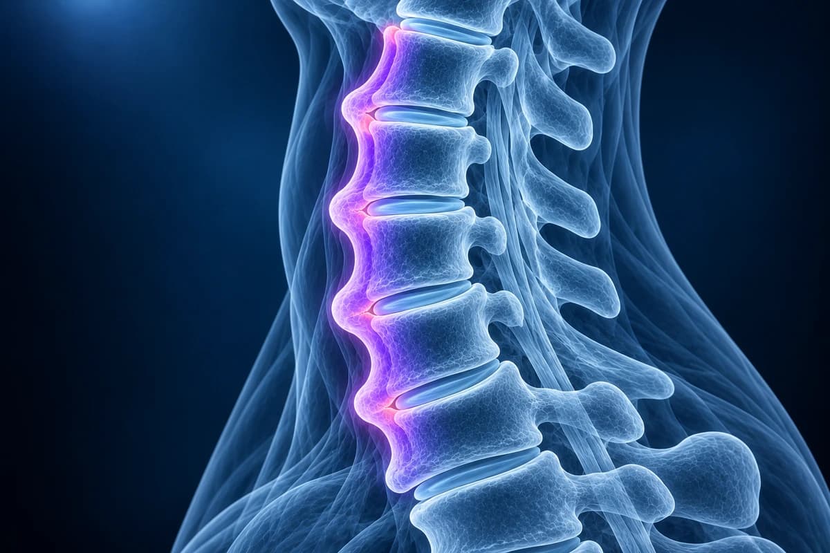
DISH (diffuse idiopathic skeletal hyperostosis) is a condition in which ligaments along the front and right side of the spine slowly turn to bone. On X-ray it is defined by flowing bridges of bone along at least four consecutive vertebrae, with disc heights largely preserved. It is often painless and found by chance, but it stiffens the spine and makes fractures after minor falls more likely.
Common Symptoms
- Morning stiffness of the neck or upper back that eases somewhat as the day goes on
- Gradual loss of neck or spine movement, often noticed when turning to look behind while driving
- Difficulty swallowing solid food, or a feeling of food sticking, when large spurs press on the gullet
- Hoarseness or a muffled voice when bony outgrowths at the front of the neck are very large
- Pain at tendon attachments such as the heel, elbow, kneecap or pelvis
- No symptoms at all, with DISH found incidentally on a chest or spine X-ray
When is neck pain with DISH an emergency?
If you have DISH and develop new neck or back pain after any fall, even from standing height or a minor bump to the head, keep your neck still and get emergency assessment. A fused, brittle spine can break like a long bone, and the break can be hard to see on plain X-ray; CT is commonly used, sometimes with MRI. Also seek urgent care for new arm or leg weakness or numbness, worsening swallowing to the point that liquids or saliva will not go down, or noisy or difficult breathing.
What does DISH mean on a spine X-ray report?
Radiologists usually use the Resnick and Niwayama criteria from 1976. All three must be present: flowing calcification or ossification along the front and side of at least four contiguous vertebral bodies, which means three or more bridged levels; relatively normal disc height without the heavy disc degeneration that would explain the bone; and no bony fusion of the facet joints or erosion and fusion of the sacroiliac joints. The last rule is what separates DISH from inflammatory spondyloarthritis.
The thoracic spine is affected most often, typically on the right side; the left is thought to be spared because the pulsating aorta lies there. The neck is also commonly involved, mostly in its lower half. Changes outside the spine are common: bony spurs where tendons and ligaments attach at the heel, the back of the elbow, the kneecap and the pelvic rim. DISH is seen mainly after the age of 50, more often in men, and has a strong link with obesity, type 2 diabetes and other features of metabolic syndrome.
What do DISH-related report phrases mean?
- "Flowing anterior osteophytes" or "bridging osteophytes"
- Thick, smooth bone running across the front of several discs and joining the vertebrae. In DISH these bridges are bulky and continuous rather than small separate beaks.
- "Ossification of the anterior longitudinal ligament"
- The ligament that lines the front of the vertebral bodies has turned into bone. Reports sometimes use the abbreviation ALL.
- "Preserved intervertebral disc spaces"
- The discs still have near-normal height. This supports DISH over severe degenerative disease, where discs collapse and spurs form at their edges.
- "Prevertebral soft-tissue impression on the oesophagus"
- Large neck spurs push the swallowing tube forward. When symptomatic this is called DISH-related dysphagia, sometimes confirmed with a swallow study.
- "Fracture through the ankylosed segment"
- A break across a fused block of spine. These injuries are often unstable because the fused segment moves as one lever, and they need specialist spine care.
For how these features look on plain films, see cervical spine X-ray explained; for the smaller, wear-related cousin of the bridging spur, see disc osteophyte complex explained.
What we look for when DISH is on the report
- The number of bridged levels and whether the bridging is continuous over four or more vertebrae
- Disc height at the bridged levels compared with the levels above and below
- Facet joints and sacroiliac joints, to separate DISH from ankylosing spondylitis
- Thickness of the bony mass in front of the neck and its relation to the airway and oesophagus
- Ossification behind the vertebral bodies (OPLL) that can narrow the spinal canal
- Any lucent line or step across a fused segment that could be a fracture
Is DISH the same as ankylosing spondylitis?
No, although both can make the spine rigid and both are sometimes called "bamboo" or "fused" spine in everyday speech. Ankylosing spondylitis is an inflammatory disease that usually starts before 45, causes inflammatory back pain that improves with exercise, inflames the sacroiliac joints early, and forms thin vertical syndesmophytes at the disc edges, with fusion of the facet joints. DISH starts later, is not primarily inflammatory, leaves the sacroiliac joint surfaces and facet joints largely intact, and forms thick, flowing bone at the front and side of the spine.
The two can occasionally occur together, and early or atypical cases can be hard to separate on a single neck film. Pelvic X-rays or sacroiliac MRI, blood tests and the overall clinical picture help a rheumatologist decide. Our spine site covers ankylosing spondylitis on its own page if you want to compare the two in more depth.
Can DISH in the neck cause swallowing or breathing problems?
Yes, occasionally. The oesophagus and windpipe sit directly in front of the lower cervical vertebrae, so a thick anterior bone mass can push on them. The most common result is difficulty swallowing solids, sometimes with hoarseness, a sense of a lump in the throat or coughing when eating. Rarely the airway is narrowed, which matters especially if a breathing tube or a camera through the throat is ever needed. Other causes of swallowing difficulty must be checked first, typically by an ENT specialist or gastroenterologist; when the bone is confirmed as the cause and symptoms are severe, surgeons can remove the anterior spurs.
Why is a fall more dangerous when you have DISH?
A normal spine absorbs a jolt across many mobile segments. A spine fused by DISH behaves like a single rigid rod, so force concentrates at one point and can snap straight through bone and ligament. A systematic review of spinal fractures in ankylosing spinal disorders, which included DISH, found these injuries were often unstable, frequently involved neurological injury and carried high complication rates; delayed diagnosis was a recurring problem. Fractures typically occur in the lower neck or at the junction between a fused and a mobile segment.
That is why emergency teams generally image the whole spine with CT, often adding MRI, when someone with DISH has new pain after even a minor fall. If you know you have DISH, it is worth telling the paramedics and the emergency doctor. Our pages on cervical spine fracture and cervical myelopathy cover the injury and cord warning signs in more detail.
Is DISH linked to OPLL and spinal stenosis in the neck?
DISH and ossification of the posterior longitudinal ligament share risk factors and often appear together. While DISH grows bone in front of the spine, where it mainly affects swallowing, OPLL grows bone behind the vertebral bodies, inside the canal, where it can squeeze the spinal cord. A report that mentions both deserves a careful look at the canal width and the cord. Ordinary cervical spondylosis can sit alongside DISH at the levels that remain mobile. Use the cervical MRI report guide for terminology accompanying DISH and canal findings.
How is DISH treated?
There is no treatment that removes the extra bone or stops it forming. Care focuses on symptoms and on the related health risks: regular stretching and mobility work, physiotherapy, pain relief as advised by your doctor, and management of weight, blood sugar, blood pressure and cholesterol, since DISH commonly travels with metabolic syndrome. Fall prevention matters more than usual. Surgery is reserved for complications such as severe swallowing difficulty, spinal cord compression from associated OPLL, or fractures.
Can I view my own spine X-ray or CT?
You can open your images privately in the free X-ray viewer or the free CT viewer and follow the front edge of the vertebrae from level to level. For a plain-language explanation of what the images and report show, upload them for an AI review. It explains findings; it is not a diagnosis and does not replace your radiologist, rheumatologist or spine surgeon.
How are imaging tests chosen for neck symptoms with DISH?
The ACR pathway quoted here applies to chronic neck pain without trauma or red flags and addresses symptoms, rather than a DISH label alone. With radiculopathy, MRI without contrast is usually appropriate; without radiculopathy, X-rays or non-contrast MRI may be appropriate.
Frequently Asked Questions
Is DISH serious?
Often it is not: many people have DISH without symptoms and learn about it from a report. It becomes important because a stiff, fused spine fractures more easily after falls, because large neck spurs can affect swallowing, and because DISH often accompanies diabetes and other metabolic risk factors worth treating.
Does DISH get worse over time?
The ossification usually progresses slowly over years, and more levels can become bridged. Staying active helps preserve movement, and follow-up imaging is usually only needed when new symptoms appear.
How many levels must be bridged to call it DISH?
Under the Resnick criteria, flowing ossification must link at least four contiguous vertebral bodies. Fewer bridged levels may be described as early or incomplete DISH, or simply as spondylosis, depending on the appearance.
Is DISH a form of arthritis?
It is grouped with arthritis-type conditions because it affects joints and ligaments, but it is not the cartilage-wearing process of osteoarthritis and not primarily an inflammatory arthritis. It is a tendency to form new bone where ligaments and tendons attach.
Will an MRI show DISH?
Yes, but X-ray and CT show the bone bridges more clearly. MRI is most useful when there is concern about the spinal cord, nerve compression, associated OPLL or a hidden fracture after injury.
Sources
- Resnick D, Niwayama G. Radiographic and pathologic features of spinal involvement in diffuse idiopathic skeletal hyperostosis (DISH). Radiology. 1976;119(3):559-568.
- Mader R, Verlaan JJ, Buskila D. Diffuse idiopathic skeletal hyperostosis: clinical features and pathogenic mechanisms. Nat Rev Rheumatol. 2013;9(12):741-750.
- Westerveld LA, Verlaan JJ, Oner FC. Spinal fractures in patients with ankylosing spinal disorders: a systematic review of the literature on treatment, neurological status and complications. Eur Spine J. 2009;18(2):145-156.
- Kuperus JS, Waalwijk JF, Regan EA, et al. Simultaneous occurrence of ankylosing spondylitis and diffuse idiopathic skeletal hyperostosis: a systematic review. Rheumatology (Oxford). 2018;57(12):2120-2128.
Understand your imaging before your next appointment
MRI, X-ray or CT from a CD, folder or ZIP. A PDF report or photos of the films work too.
Free to open and preview, no account needed. An optional AI report from $10 explains your images in plain language, with questions for your doctor.
Your raw files stay on this device. Only rendered images and minimal context are sent when you request an AI report.
Medical Disclaimer: This page is for informational and educational purposes only. It does not constitute medical advice, diagnosis, or treatment. AI-generated analysis may contain errors. Always consult a qualified healthcare professional for medical decisions. Full Disclaimer