
Disc Osteophyte Complex in the Neck: What Your MRI Report Means
A plain-language guide to "disc osteophyte complex" on a cervical MRI: what the ridge is made of, how it differs from a herniated disc, and what effacing the thecal sac, cord contact and cord compression mean.
Key Takeaways
- A disc osteophyte complex is a combined ridge of bulging disc and bone spurs from age-related wear
- The words after it matter most: indenting the sac, contacting the cord or compressing the cord
- Disc bulging is seen in most adults on MRI; cord compression and cord signal change are much rarer
- Clumsy hands, walking changes or worsening weakness need prompt review whatever the report says
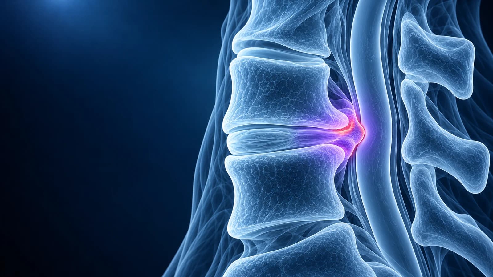
A “disc osteophyte complex” is a ridge at the back of a neck disc space made of two things that sit together: the bulging edge of a worn disc and the bony spurs (osteophytes) that grow from the rims of the vertebrae above and below it.
On MRI the two are hard to tell apart, so the report names them as one. It is a sign of age-related wear, not an injury. What matters is the next part of the sentence: whether the ridge only indents the fluid around the spinal cord, touches the cord, or flattens it, and whether it narrows the holes where the nerves leave.
This guide explains how the ridge forms, how it differs from a herniated disc, how to read the severity words that follow it, and when a finding like this needs prompt attention. It is educational and does not replace review by the clinician who knows your symptoms and examination.
How does a disc osteophyte complex form?
With age, the discs between neck vertebrae lose water and height. A thinner disc bulges outward in a broad band, rather like a tyre with low pressure, and the bone rims next to it take more load. The body responds by growing bony lips along those rims. Over years, the bulge and the spurs fuse into a firm ridge running across the back of the disc space. This process is part of cervical spondylosis and is most often reported at C5–C6 and C6–C7, the levels that bend the most in daily life.
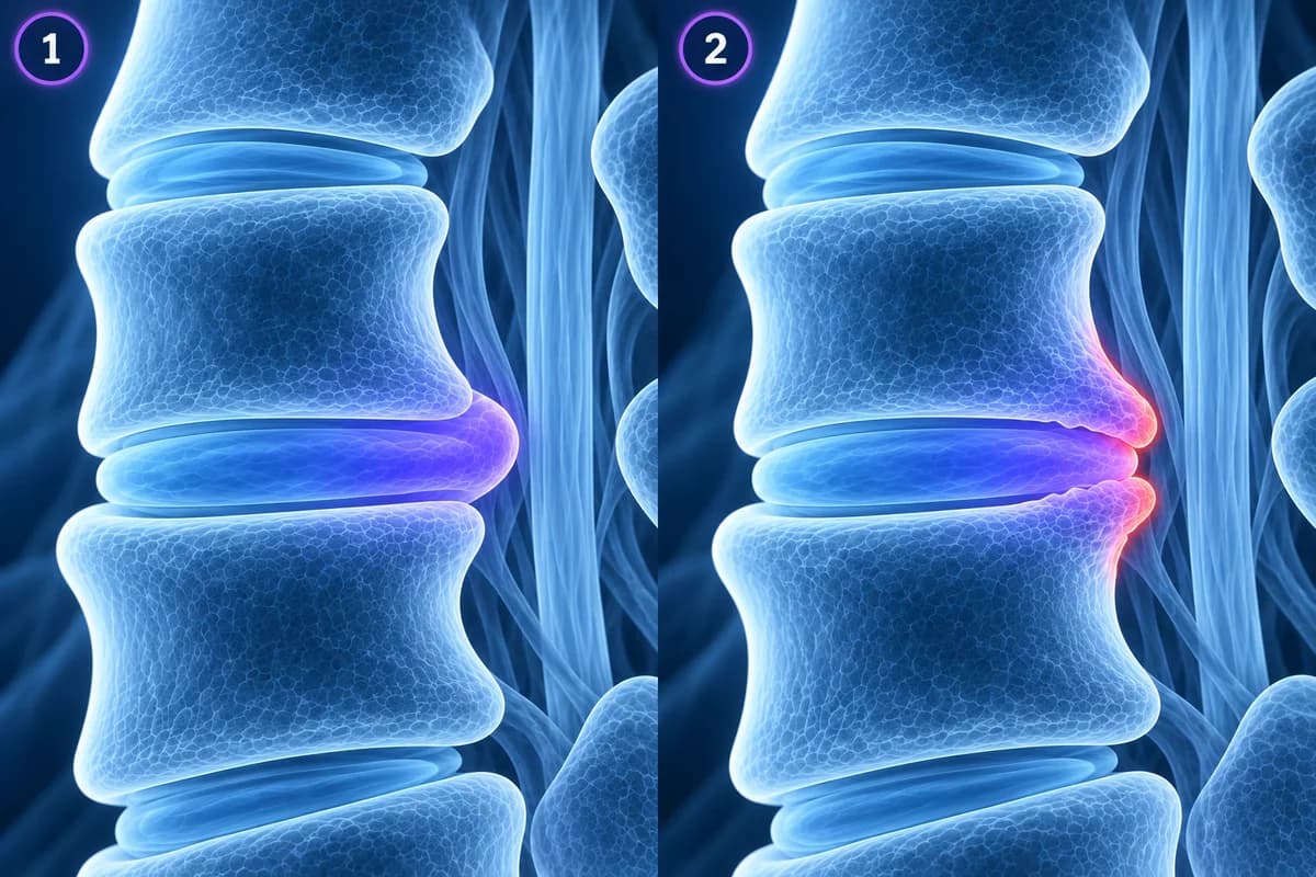
Surgeons sometimes call this a “hard disc”, in contrast with a “soft disc” herniation of fresh disc material. The distinction matters because a soft fragment can shrink by itself over months, while the bony part of a ridge does not go away.
Is a disc osteophyte complex the same as a herniated disc?
Not quite. A herniation is disc material pushed beyond its normal edge in a focal spot, often appearing over days or weeks and often causing sudden arm pain. A disc osteophyte complex is broader, built up slowly and partly made of bone. The two can coexist, and some reports use “disc osteophyte complex with superimposed protrusion” to say a newer soft bulge sits on top of an older ridge. If your report says “protrusion” or “extrusion” instead, our pages on cervical disc herniation and C5–C6 and C6–C7 herniations are the better fit.
Some radiologists dislike the term because it hides the mix of disc and bone. When the bony share matters, for instance before surgery, a CT scan shows the spurs much more clearly than MRI. CT also separates the ridge from an ossified ligament running behind several vertebrae, a different condition called OPLL. Our comparison of CT and MRI for cervical radiculopathy explains what each test adds.
What does “effacing the thecal sac” mean, and how severe is it?
The spinal cord floats in a sleeve of fluid (cerebrospinal fluid, or CSF) inside a membrane called the thecal sac. On T2-weighted MRI that fluid is bright, framing the darker cord. The verbs in your report describe how far the ridge has pushed into this space, and they roughly form a ladder:
- “Indenting” or “mildly effacing the ventral thecal sac”: the ridge presses on the front of the fluid sleeve but fluid still surrounds the cord. Very common, often painless.
- “Effacing the ventral CSF” or “obliterating the anterior subarachnoid space”: the fluid in front of the cord is squeezed out at that level, but the cord keeps its shape.
- “Abutting” or “contacting the cord”: the ridge touches the cord surface without reshaping it.
- “Flattening”, “deforming” or “compressing the cord”: the cord itself is pushed out of its round or oval shape. This is cord compression and is reported with care.
- “With T2 hyperintense cord signal”: a bright spot inside the cord at that level, which can mean the cord is irritated or injured by the pressure.
A grading system published in 2011 formalised this ladder for neck MRI: grade 0, no narrowing; grade 1, more than half of the fluid space obliterated without the cord changing shape; grade 2, cord deformity; grade 3, signal change inside the cord. Six radiologists using it agreed well on whether a level was grade 0–1 or 2–3. Many reports still use the words “mild”, “moderate” and “severe” instead; ask which rung of the ladder they mean if the report is unclear. The signs that the cord itself is affected are covered in cervical myelopathy warning signs on MRI.
How common is a disc osteophyte complex in people without symptoms?
Very common at the milder end. A Japanese study scanned the necks of 1,211 healthy volunteers aged 20 to 70. Disc bulging was seen in 87.6% of them, including most people in their twenties, and it grew more frequent and more pronounced with each decade. Actual spinal cord compression was much rarer, at 5.3%, and a bright cord signal appeared in 2.3%, both becoming more frequent after age 50. In other words, a ridge that indents the sac is an expected finding in many adults, while a ridge that deforms the cord is unusual enough to be matched carefully against symptoms and examination.
What does it mean if the ridge points into the foramen?
The report usually says where the ridge is widest: “central” (toward the cord), “paracentral” (to one side of the cord) or “foraminal” (into the side opening where a nerve root exits). Spurs from the small uncovertebral joints at the side edges of each disc, written as “uncovertebral hypertrophy” or “uncinate spurring”, often join the ridge and narrow the foramen. When that narrowing pinches a root, it can cause pain, tingling or weakness in one arm in a pattern that matches the level, which is cervical radiculopathy. The narrowing itself has its own page: cervical foraminal stenosis.
Which scan shows a disc osteophyte complex best?
- X-ray: shows lower disc heights and bony lips at the vertebral corners, but not the cord or the nerves.
- MRI: shows the ridge, the fluid around the cord, the cord shape and cord signal, and the nerve exits. The American College of Radiology rates MRI without contrast as usually appropriate for new or increasing arm symptoms from the neck.
- CT: shows exactly how much of the ridge is bone, which helps surgical planning and separates spurs from an ossified ligament. It shows the cord much less well.
Does a disc osteophyte complex need surgery?
Most do not. Neck pain and many episodes of arm pain linked to spondylosis improve with time, activity adjustment, physiotherapy and pain relief. Surgery is considered when the ridge compresses the cord and there are signs of myelopathy, or when a pinched nerve causes persistent or worsening weakness or pain that does not settle with non-surgical care. The usual operation removes the ridge from the front and fuses the level; what imaging looks for afterwards is explained in cervical MRI after fusion. These decisions belong to a spine specialist who has examined you.
Which symptoms with a disc osteophyte complex need urgent care?
Get prompt medical review if you notice clumsy hands (dropping things, trouble with buttons or writing), numbness in both hands, a change in balance or walking, arm or leg weakness that is getting worse, or new bladder or bowel problems. These can be signs that the cord is affected. Neck pain after a fall or collision, fever with neck pain, or a history of cancer or a weakened immune system also needs same-day assessment.
How do I find the ridge on my own MRI?
Load your DICOM files into our free MRI viewer, which opens them in your browser without uploading the originals. Pick the sagittal T2 series, find the disc level the report names by counting down from C2, and look at the bright fluid in front of the cord. Then switch to the axial T2 series at the same level to see whether the cord stays oval or is flattened, and whether each foramen is open. For the other phrases on the same report, see cervical MRI report phrases explained and how to read a cervical spine MRI.
Frequently Asked Questions
Is a disc osteophyte complex serious?
It depends on what it touches. A ridge that only indents the thecal sac is common and often causes no symptoms. A ridge that flattens the cord, causes a cord signal change or pinches a nerve root that matches your symptoms deserves specialist attention.
Can a disc osteophyte complex shrink or go away?
The bony part does not shrink. A soft disc component on top of it can sometimes settle, and symptoms often improve even when the ridge looks the same on a later scan, because pain depends on more than the shape.
What is the difference between a disc bulge and a disc osteophyte complex?
A disc bulge is soft disc tissue extending broadly beyond the vertebral edges. A disc osteophyte complex adds bone spurs from the vertebral rims, so the ridge is firmer and partly bony. Reports often use the second term in older necks where both are present.
Can a disc osteophyte complex cause headaches?
Ridges at the lower neck levels mainly affect the arm nerves or the cord. Headache that starts in the neck is more often linked to the upper joints, as described under cervicogenic headache. A severe sudden headache with a stiff neck is an emergency.
Can AI read the disc osteophyte complex on my MRI?
AI can point out the level, explain the severity words and show where the ridge sits on your images. It is not a radiologist, and decisions about treatment need a clinician who has examined your hands, arms and walking.
Sources
- Kang Y, Lee JW, Koh YH, et al. New MRI grading system for the cervical canal stenosis. AJR Am J Roentgenol. 2011;197(1):W134-W140.
- Nakashima H, Yukawa Y, Suda K, et al. Abnormal findings on magnetic resonance images of the cervical spines in 1211 asymptomatic subjects. Spine (Phila Pa 1976). 2015;40(6):392-398.
- Margetis K, Tadi P. Cervical Spondylosis. StatPearls. Treasure Island (FL): StatPearls Publishing.
- Expert Panel on Neurological Imaging; McDonald MA, Kirsch CFE, et al. ACR Appropriateness Criteria Cervical Neck Pain or Cervical Radiculopathy. J Am Coll Radiol. 2019;16(5S):S57-S76.
Related Articles
What your neck MRI report means: uncovertebral hypertrophy, foraminal narrowing grades, effacement of the CSF, cord flattening, T2 hyperintense cord signal, Modic changes and more, each explained in one or two plain sentences.
MRI is usually the first scan for a pinched nerve in the neck, while CT shows bone spurs, OPLL and foraminal narrowing in finer detail. Learn when each is used, when CT myelography is needed, and what the report phrases mean.
A plain-language guide to your neck MRI: how to count from C2 down to C7, which slices show the cord and the nerve exits, and which artifacts make a cervical scan look worse than it is.
Related Conditions
Understand your imaging before your next appointment
MRI, X-ray or CT from a CD, folder or ZIP. A PDF report or photos of the films work too.
Free to open and preview, no account needed. An optional AI report from $10 explains your images in plain language, with questions for your doctor.
Your raw files stay on this device. Only rendered images and minimal context are sent when you request an AI report.
Medical Disclaimer: This page is for informational and educational purposes only. It does not constitute medical advice, diagnosis, or treatment. AI-generated analysis may contain errors. Always consult a qualified healthcare professional for medical decisions. Full Disclaimer