
Cervical Radiculopathy: Matching Your MRI Level to Your Arm Symptoms
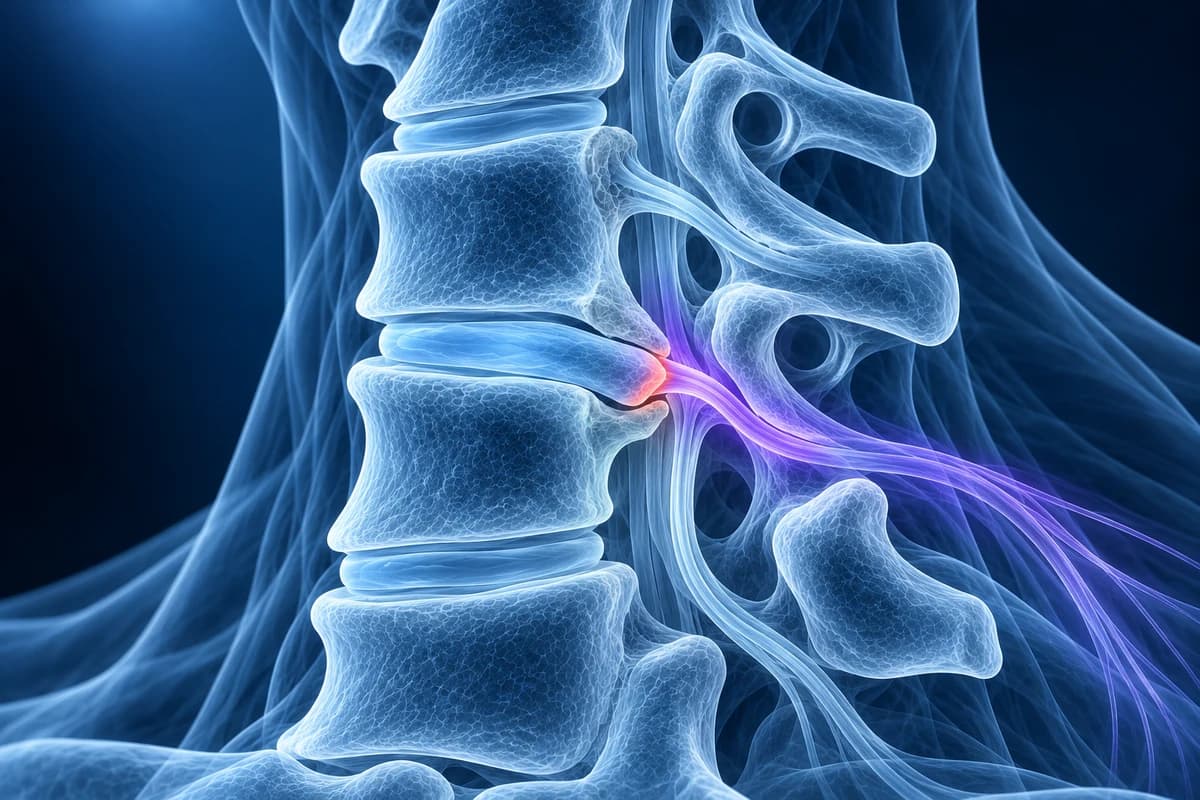
Cervical radiculopathy is irritation or compression of a nerve root as it leaves the neck, causing pain, tingling or weakness in one arm. On MRI the key is matching level to symptoms: a problem at C5–C6 usually affects the C6 root (thumb side of the forearm and hand), and C6–C7 usually affects the C7 root (middle finger and triceps). Most cases settle without surgery.
Common Symptoms
- Sharp, burning or electric pain that travels from the neck into the shoulder blade, arm or hand on one side
- Pins and needles or numbness in a specific strip of the arm, such as the thumb and index finger or the middle finger
- Weakness in one movement, for example lifting the arm sideways, bending the wrist back or straightening the elbow
- Arm pain that gets worse when tilting the head back and turning it toward the painful side
- Relief when resting the hand of the painful arm on top of the head
- A reduced biceps, brachioradialis or triceps reflex found by the examining clinician
When does a pinched nerve in the neck need urgent care?
Seek same-day medical assessment if arm or hand weakness is getting worse over hours or days, if both arms are affected, if your hands have become clumsy (dropping things, trouble with buttons or writing), if your walking or balance has changed, or if you notice new bladder or bowel problems. These suggest the spinal cord itself may be compressed, which is a different and more urgent problem called cervical myelopathy. Neck pain after a fall or car crash, neck pain with fever, or arm pain in someone with a history of cancer or a weakened immune system also needs prompt review rather than waiting for a scan explanation.
Why is the nerve numbered differently from the disc level?
The neck has seven vertebrae but eight pairs of cervical nerve roots. Each cervical root leaves the canal above the vertebra that shares its number: the C6 root exits between C5 and C6, the C7 root between C6 and C7, and the C8 root between C7 and the first thoracic vertebra. That is why a report describing a problem "at C5–C6" is usually linked to C6 symptoms, not C5 symptoms. People reading their own MRI often look for the wrong nerve because of this offset.
The most frequently affected roots are C7 and C6, because the C5–C6 and C6–C7 levels carry the most movement and wear in the neck. The rough pattern clinicians use to match symptoms to a root is:
- C5 root (C4–C5 level): outer shoulder and upper arm, weakness lifting the arm out to the side.
- C6 root (C5–C6 level): outer forearm, thumb and index finger, weakness bending the elbow or lifting the wrist, reduced biceps or brachioradialis reflex.
- C7 root (C6–C7 level): back of the arm into the middle finger, weakness straightening the elbow, reduced triceps reflex.
- C8 root (C7–T1 level): inner forearm, ring and little fingers, weakness of grip and finger movements.
These maps overlap from person to person, so they guide rather than prove. Our explainer on C5–C6 and C6–C7 disc herniations goes through the two busiest levels in more detail.
What does a cervical radiculopathy MRI report actually say?
- "Left foraminal narrowing at C6–C7"
- The side opening (foramen) where the left C7 root exits is smaller than usual. Side matters: left-sided narrowing should match left arm symptoms. Reports usually grade it as mild, moderate or severe.
- "Contacts", "abuts" or "impinges on the exiting nerve root"
- The radiologist is describing how close the disc or bone is to the nerve. "Contacts" and "abuts" mean touching; "impinges" or "compresses" means the root is pushed or flattened. Touching alone does not always cause symptoms.
- "Uncovertebral hypertrophy" or "uncovertebral joint spurring"
- The small joints at the front corners of each cervical vertebra have grown bony edges with wear. They form the front wall of the nerve opening, so this is one of the commonest causes of a pinched cervical root in people over about 40.
- "Disc osteophyte complex"
- A combined ridge of bulging disc and bone spur at the back of the disc space. MRI cannot always separate the soft disc part from the bony part, so the two are reported together. See our page on the disc osteophyte complex.
- "Posterolateral" or "foraminal disc protrusion"
- Soft disc material has pushed backward and to one side, toward the nerve opening rather than the centre of the canal. This shape is typical of the sudden, younger-patient form of radiculopathy covered on our cervical disc herniation page.
More wording is decoded in cervical MRI report phrases explained and in our step-by-step guide on how to read a cervical spine MRI.
What causes a pinched nerve in the neck: soft disc or bone spur?
There are two broad patterns. In people in their thirties and forties, a sudden onset of arm pain after lifting, sleeping awkwardly or for no clear reason is often caused by a soft disc protrusion pressing on the root inside or just at the entrance of the foramen. Soft disc fragments can shrink on their own over weeks to months, which is one reason the outlook is often good.
In older adults, the cause is more often a gradual narrowing of the nerve opening by uncovertebral and facet joint spurs, a stiffened disc and a lower disc height. This is the same wear process described on our cervical spondylosis page, and when the opening itself is the main problem it is reported as cervical foraminal stenosis. Symptoms from bony narrowing tend to come and go with posture and activity rather than appearing all at once. Less common causes include nerve sheath tumours, infection and inflammatory disease, which is why unusual features prompt further tests.
Do you need an MRI for cervical radiculopathy?
Not always straight away. Many people with a first episode of arm pain and no warning signs are managed with activity changes, pain relief and physiotherapy first, because the condition often improves on its own. Imaging is usually added when pain persists beyond several weeks, when there is measurable weakness, when the diagnosis is uncertain, or when an injection or operation is being considered.
The ACR Appropriateness Criteria for cervical neck pain or radiculopathy list MRI of the cervical spine without contrast and plain X-rays among the appropriate first studies for new or worsening radiculopathy. MRI shows discs, nerve roots and the spinal cord. X-rays show alignment, disc height and large spurs; oblique views can show the bony outline of the foramina. CT shows bone edges sharply and is useful when the question is whether a spur or a soft disc is doing the pinching, while CT myelography is used when MRI is not possible or is inconclusive. Our comparison of CT vs MRI for cervical radiculopathy and the guide do you need an MRI for neck pain cover the choice in more depth.
Nerve conduction studies and electromyography (EMG) are sometimes added to separate a neck root problem from a trapped nerve at the wrist or elbow, such as carpal or cubital tunnel syndrome, which can produce similar hand numbness.
What we look for on cervical radiculopathy imaging
- The level and side of foraminal narrowing, checked against the arm and fingers you describe
- Posterolateral or foraminal disc protrusions and extrusions that reach the exiting nerve root
- Uncovertebral and facet joint spurs narrowing the nerve opening on axial images
- Loss of disc height and disc osteophyte complexes at C5–C6 and C6–C7
- Central canal narrowing or cord contact that would change the urgency of the picture
- Unusual findings such as a nerve sheath mass, bone marrow replacement or signs of infection
Why does my MRI show narrowing on the side that does not hurt?
Foraminal narrowing and disc bulges are very common on neck MRIs of people with no arm symptoms at all, and they become more common with age. A report may list several levels and both sides. The finding that matters is the one whose level and side fit your symptoms and examination. A clinician weighs the report against where the pain travels, which muscles are weak and which reflexes are reduced. When those do not line up, they may look for another cause rather than treat the most dramatic-looking level.
How is cervical radiculopathy treated, and when is surgery considered?
Most people improve over weeks to a few months with non-operative care: short-term pain medication, guided exercise and physiotherapy, posture and sleep adjustments, and sometimes a short course of oral steroids prescribed by a clinician. A targeted epidural or nerve root injection can be offered for severe arm pain that is not settling.
Surgery is usually discussed when arm pain stays disabling despite a fair trial of these measures, when weakness is significant or progressing, or when cord compression is also present. Common options are anterior cervical discectomy and fusion (ACDF), cervical disc replacement, or a posterior foraminotomy that widens the nerve opening from behind. If you have already had surgery and have new symptoms, see cervical MRI after fusion. The right choice depends on your images, your examination and your goals, and is made with a spine surgeon.
A similar nerve root problem in the lower back causes sciatica down the leg. That is a separate topic covered by lumbar spine resources, not on this neck site.
Can I look at my own neck MRI for a pinched nerve?
Yes. Open your DICOM files privately in the free cervical MRI viewer and step through the axial slices at the level your report names to compare the left and right nerve openings. If you had CT, the free CT viewer shows the bone spurs more clearly. For a plain-language walk-through of your images and report, upload them for an AI review. It explains findings; it is not a diagnosis and does not replace your radiologist, GP or spine specialist.
Frequently Asked Questions
Which nerve is affected by a C5–C6 disc problem?
Usually the C6 nerve root, because cervical roots exit above the vertebra with the same number. C6 symptoms typically involve the outer forearm, thumb and index finger, with weakness bending the elbow or lifting the wrist. A large central disc at C5–C6 can also press on the spinal cord, which is assessed separately.
How long does cervical radiculopathy take to get better?
Many people notice clear improvement within weeks to a few months of non-operative care, although numbness and mild weakness can take longer to settle than pain. Symptoms that are worsening, or weakness that is not improving, should be reviewed rather than waited out.
Can a pinched nerve in the neck cause hand numbness?
Yes. C6 problems often cause thumb and index finger numbness, C7 the middle finger and C8 the ring and little fingers. Carpal tunnel and cubital tunnel syndrome can produce similar patterns, so a clinician may test sensation, strength and reflexes, or order nerve studies, to find where the nerve is being squeezed.
Is foraminal narrowing on my MRI the same as radiculopathy?
No. Foraminal narrowing is an imaging finding; radiculopathy is a clinical diagnosis based on symptoms and examination. Many people have narrowing without symptoms. The two are linked only when the level and side of the narrowing match the arm symptoms.
Should I see a doctor if only my arm hurts and my neck feels fine?
Yes. Radiculopathy often causes more arm pain than neck pain. A clinician can check strength and reflexes, which tells them how much the nerve is affected and whether imaging is needed now. Any weakness, clumsy hands or change in walking should be seen promptly.
Sources
- Expert Panel on Neurological Imaging; McDonald MA, Kirsch CFE, Amin BY, et al. ACR Appropriateness Criteria Cervical Neck Pain or Cervical Radiculopathy. J Am Coll Radiol. 2019;16(5S):S57-S76.
- Margetis K, Magnus W, Mesfin FB. Cervical Radiculopathy. StatPearls. Treasure Island (FL): StatPearls Publishing.
- American Academy of Orthopaedic Surgeons. Cervical Radiculopathy (Pinched Nerve). OrthoInfo.
Related Articles
Why C5-C6 and C6-C7 are the neck discs that herniate most, which nerve root each one pinches, how the arm symptoms differ, and what protrusion, extrusion and foraminal mean on your MRI.
MRI is usually the first scan for a pinched nerve in the neck, while CT shows bone spurs, OPLL and foraminal narrowing in finer detail. Learn when each is used, when CT myelography is needed, and what the report phrases mean.
What an MRI can and cannot show after an anterior cervical fusion (ACDF): metal artifact, adjacent segment disease, residual compression and cord signal, when CT or X-ray answers better, and which symptoms need urgent care.
Understand your imaging before your next appointment
MRI, X-ray or CT from a CD, folder or ZIP. A PDF report or photos of the films work too.
Free to open and preview, no account needed. An optional AI report from $10 explains your images in plain language, with questions for your doctor.
Your raw files stay on this device. Only rendered images and minimal context are sent when you request an AI report.
Medical Disclaimer: This page is for informational and educational purposes only. It does not constitute medical advice, diagnosis, or treatment. AI-generated analysis may contain errors. Always consult a qualified healthcare professional for medical decisions. Full Disclaimer