
Atlantoaxial Instability: Measuring the C1–C2 Gap and When It Matters
Atlantoaxial instability means excess movement between the first two neck vertebrae, the atlas (C1) and the axis (C2). On a side X-ray it is measured as the atlanto-dens interval, the gap between the front ring of C1 and the peg of C2: above about 3 mm in adults or about 5 mm in children is generally considered abnormal. It is most often seen in rheumatoid arthritis, Down syndrome and after trauma.
Some signs described on this page need prompt medical care. If you have them, get care first. Reading about imaging can wait.
Common Symptoms
- Pain high in the neck or at the base of the skull, often worse when nodding the head forward
- A head that tilts to one side or a neck that resists turning, especially in children
- Electric-shock sensations down the spine or into the limbs when bending the neck forward
- Clumsy hands, dropping objects, or a new unsteady or wide-based walk
- In children with Down syndrome, losing skills such as walking, new tiredness when walking, or a change in toileting
- Many people, particularly those found on routine X-rays, have no symptoms at all
Which symptoms need same-day care?
Seek urgent medical assessment if someone with known or suspected C1–C2 instability develops new weakness, numbness or tingling in the arms or legs, clumsy hands, a change in walking or balance, loss of bladder or bowel control, electric shocks down the spine when bending the head, or episodes of dizziness, fainting, double vision or difficulty swallowing. In a child with Down syndrome, a sudden head tilt, neck pain, refusal to walk or loss of a skill they had mastered deserves the same urgency. After any neck injury in these groups, keep the neck still and get emergency care. This page explains imaging findings; it does not diagnose or clear anyone.
How does the C1–C2 joint normally work?
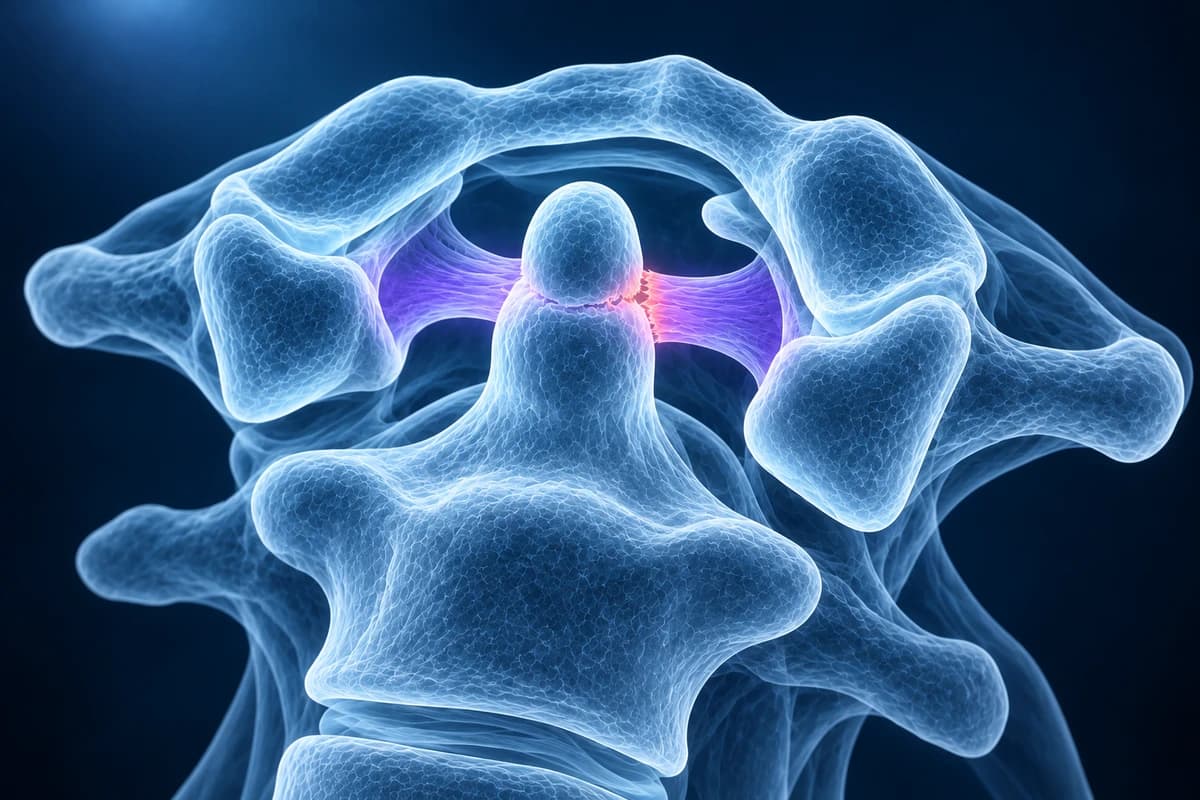
Unlike the lower neck, C1 and C2 have no disc between them. The atlas is a ring, and the axis sends a bony peg, the dens or odontoid process, up through the front of that ring. The transverse ligament straps the dens against the back of the front arch of C1, and the alar ligaments tie the tip of the dens to the skull. This arrangement lets the head rotate: roughly half of all neck rotation happens at this single level. The trade-off is that stability depends almost entirely on ligaments rather than bone shape.
When the transverse ligament is stretched, eroded or torn, C1 can slide forward on C2 as the head nods. The dens then moves backward relative to the ring, narrowing the space available for the spinal cord at the very top of the neck. Because the cord here controls breathing as well as all four limbs, severe compression at C1–C2 is dangerous, which is why clinicians take this measurement seriously even when symptoms are mild.
What is the atlanto-dens interval and what is normal?
The anterior atlanto-dens interval (ADI, also called the atlanto-dental interval) is measured on a lateral neck X-ray or a sagittal CT from the back edge of the front arch of C1 to the front edge of the dens. In adults it is normally 3 mm or less. Children have more flexible ligaments and the dens is still partly cartilage, so up to about 5 mm is accepted in young children. The number matters most when it changes between positions: a gap that widens when the head bends forward is the real sign of instability.
The posterior atlanto-dens interval (PADI), sometimes called the space available for the cord, is measured from the back of the dens to the front of the C1 back arch. In a long-term study of rheumatoid arthritis patients, a PADI below 14 mm was associated with a higher risk of paralysis, and many surgeons now weigh the PADI more heavily than the anterior gap. MRI adds a direct view of the cord and of any soft tissue around the dens.
What does a flexion-extension X-ray show at C1–C2?
A flexion-extension study is a pair of side X-rays taken with the chin tucked down and then with the head tilted back. The radiologist measures the atlanto-dens interval on both. In instability the gap opens in flexion and closes in extension. These views are taken with the patient moving actively and only as far as comfortable, never forced, and they are not used in acute trauma until a fracture has been excluded by CT. Our flexion-extension X-ray guide explains how the views are performed and what other levels are checked, and the cervical spine X-ray explainer covers the standard side view.
MRI, sometimes performed in flexion and extension, shows whether the cord is compressed and whether it has abnormal signal. CT shows the bony anatomy in detail and is the test for rotatory problems and congenital variants. After an injury, the ACR Appropriateness Criteria for suspected spine trauma guide which combination is appropriate.
Why does rheumatoid arthritis affect C1–C2?
The joints around the dens are lined with synovium, the same tissue rheumatoid arthritis attacks in the hands and feet. Inflamed synovium, called pannus, erodes the dens and loosens the transverse ligament. The result can be anterior atlantoaxial subluxation, the most common neck problem in long-standing rheumatoid arthritis. Over time some patients also develop cranial settling, where the skull sinks onto C1 and the dens rises toward the base of the brain, and subaxial subluxations lower down that give a stepladder appearance on X-ray.
Modern drug treatment has made severe cervical involvement less common, but rheumatology and anaesthesia teams still ask about neck symptoms before surgery, because intubation flexes the neck. A report phrase such as "pannus formation around the dens" or "erosion of the odontoid process" means inflammatory tissue and bone loss at this joint.
Should children with Down syndrome have neck X-rays?
Ligament laxity in Down syndrome makes a widened atlanto-dens interval more common, but most children with a widened gap never develop cord problems, and a normal X-ray does not guarantee that problems will not develop later. For that reason, the American Academy of Pediatrics no longer recommends routine screening X-rays in children without symptoms. Instead, parents and clinicians watch for warning signs (neck pain, head tilt, change in walking, weakness, loss of skills, change in bladder or bowel function) and image promptly if they appear, usually starting with neutral lateral X-rays and moving to flexion-extension views and MRI.
Families are often advised to discuss neck positioning before anaesthesia and before sports with a risk of neck flexion or head impact. Individual decisions belong to the child's paediatrician or spine specialist.
What do C1–C2 report phrases mean?
- "Widened atlantodental interval"
- The gap in front of the dens is larger than expected for age. Reports usually give the number in millimetres and, on a dynamic study, the value in each position.
- "Atlantoaxial subluxation"
- C1 has shifted partly out of its normal position on C2. Anterior subluxation is the most common direction; rotatory subluxation, where C1 is locked in a turned position, mainly occurs in children.
- "Os odontoideum"
- The top of the dens is a separate, smooth-edged bone not fused to C2. It may be developmental or the result of an old injury, and it can make C1–C2 unstable.
- "Basilar invagination" or "cranial settling"
- The tip of the dens sits higher than normal relative to the skull base, which can press on the brainstem. It is seen in advanced rheumatoid arthritis and in some congenital conditions.
- "Cord compression at C1–C2" with "T2 hyperintense cord signal"
- MRI shows the cord narrowed at the top of the neck, with a bright area that suggests swelling or injury to the cord tissue. This finding usually prompts a surgical opinion.
More wording is explained in the cervical MRI report phrases guide, and the signs of cord involvement are covered in cervical myelopathy warning signs on MRI.
What we look for at the craniocervical junction
- Anterior atlanto-dens interval in neutral, flexion and extension, compared with age-appropriate limits
- Posterior atlanto-dens interval and remaining space available for the cord
- Dens erosion, pannus, os odontoideum or an old odontoid fracture
- Position of the dens tip relative to the skull base for basilar invagination
- Rotational asymmetry of the C1 lateral masses on C2
- Cord narrowing or T2 hyperintense cord signal at the C1–C2 level on MRI
What else causes C1–C2 instability, and how is it treated?
Trauma can rupture the transverse ligament, sometimes alongside a C1 ring fracture; that pathway is explained on our cervical spine fracture page. Infection of the throat or upper neck in children can loosen the ligaments and cause a painful rotatory subluxation, and infections of the spine itself are covered under discitis and osteomyelitis. Rarer causes include skeletal dysplasias and connective tissue disorders.
People without symptoms and with modest widening are usually monitored with periodic examination and imaging, while avoiding activities with a high risk of neck impact. Progressive widening, a small space for the cord, cord signal change or myelopathy symptoms usually lead to surgery that fuses C1 to C2 with screws, sometimes extending to the skull. A spine surgeon makes that decision with the full set of images and the clinical examination. Lower neck cord compression from wear is a separate problem, described on the cervical myelopathy page.
Can I measure the C1–C2 gap on my own X-ray?
You can open your lateral and flexion-extension films privately in the free neck X-ray viewer and use its measuring tool to see the gap your report describes, or review an MRI in the free MRI viewer. Files never leave your device. For a plain-language explanation of your report, upload the images for an AI review. Home measurements are not precise enough to make decisions; they are a way to follow the conversation with your doctor, not a diagnosis.
Frequently Asked Questions
What is a normal atlanto-dens interval?
In adults the anterior atlanto-dens interval is normally 3 mm or less. In young children up to about 5 mm is accepted because their ligaments are looser. The measurement is more meaningful when compared between flexion and extension and alongside the space left for the spinal cord behind the dens.
Is atlantoaxial instability dangerous?
It can be, because the spinal cord at C1–C2 controls breathing and all four limbs. Many people with mild widening never develop problems, but symptoms such as clumsy hands, gait change or electric shocks with neck bending need prompt assessment, and neck trauma in a person with known instability is an emergency.
Does atlantoaxial instability always need surgery?
No. Asymptomatic people with stable, modest widening are usually monitored. Surgery, typically a C1–C2 fusion, is considered when the gap keeps increasing, when the space for the cord becomes small, when MRI shows cord damage, or when there are neurological symptoms.
Can a flexion-extension X-ray be harmful?
When ordered appropriately, the movement is performed by the patient within a comfortable range, so the risk is low. The views are not used straight after significant trauma until a fracture has been excluded, and some clinicians prefer MRI or supervised imaging in people with severe instability or cord symptoms.
Is atlantoaxial instability the same as an odontoid fracture?
No, although a fracture of the dens can make C1–C2 unstable. Atlantoaxial instability usually refers to ligament weakness or damage that lets C1 slide on an intact C2. An odontoid fracture is a break in the bone and is managed as an acute fracture.
Sources
- Lacy J, Bajaj J, Gillis CC. Atlantoaxial Instability. StatPearls. Treasure Island (FL): StatPearls Publishing.
- Boden SD, Dodge LD, Bohlman HH, Rechtine GR. Rheumatoid arthritis of the cervical spine. A long-term analysis with predictors of paralysis and recovery. J Bone Joint Surg Am. 1993;75(9):1282-1297.
- Bull MJ, Trotter T, Santoro SL, et al. Health supervision for children and adolescents with Down syndrome. Pediatrics. 2022;149(5):e2022057010.
- Expert Panel on Neurological Imaging and Musculoskeletal Imaging; Beckmann NM, West OC, et al. ACR Appropriateness Criteria Suspected Spine Trauma. J Am Coll Radiol. 2019;16(5S):S264-S285.
Related Articles
Why flexion and extension neck X-rays are ordered, how the test is done safely, which measurements suggest instability, why they are not the first test after an injury, and what the report wording means.
What a neck X-ray can and cannot show: the standard views, the four alignment lines, disc space narrowing, bone spurs and the Torg-Pavlov ratio, and when CT or MRI is needed instead.
Do not delay urgent medical care to use this tool.
After you get care, understand your imaging
MRI, X-ray or CT from a CD, folder or ZIP. A PDF report or photos of the films work too.
Free to open and preview, no account needed. An optional AI report from $10 explains your images in plain language, with questions for your doctor.
Your raw files stay on this device. Only rendered images and minimal context are sent when you request an AI report.
Medical Disclaimer: This page is for informational and educational purposes only. It does not constitute medical advice, diagnosis, or treatment. AI-generated analysis may contain errors. Always consult a qualified healthcare professional for medical decisions. Full Disclaimer