
Cervical Myelopathy: Spinal Cord Compression and What Your MRI Shows
Cervical myelopathy is dysfunction of the spinal cord caused by compression in the neck, most often from age-related disc, bone and ligament changes (degenerative cervical myelopathy). The typical signs are clumsy hands and unsteady walking. On MRI, the cord looks squeezed, and a bright spot inside it on T2 images ("T2 hyperintense cord signal") suggests the cord itself is affected.
Some signs described on this page need prompt medical care. If you have them, get care first. Reading about imaging can wait.
Common Symptoms
- Clumsy hands: dropping objects, fumbling with buttons, keys or coins, or handwriting getting worse
- Numbness or a "wearing gloves" feeling in both hands that does not follow a single finger pattern
- Unsteady, wide-based walking, tripping, or needing to hold the rail on stairs
- An electric shock running down the spine or into the limbs when bending the head forward
- Stiff or heavy legs, and brisk reflexes or a positive Hoffmann sign found on examination
- Bladder urgency, frequency or difficulty passing urine, usually in more advanced cases
Which myelopathy symptoms need same-day care?
Go to an emergency department or call emergency services if you have new or rapidly worsening weakness in the arms or legs, you are suddenly unable to walk safely, you lose control of your bladder or bowels, or you develop numbness around the trunk. The same applies after any fall or neck injury in someone already known to have a narrow canal, because a minor injury can bruise an already compressed cord. Slowly progressive symptoms, such as hands getting clumsier over months or a gradual change in balance, are not an emergency, but they do need prompt referral to a spine specialist rather than waiting to see if they pass. Neck pain with fever, or with a history of cancer or a weakened immune system, also needs urgent review.
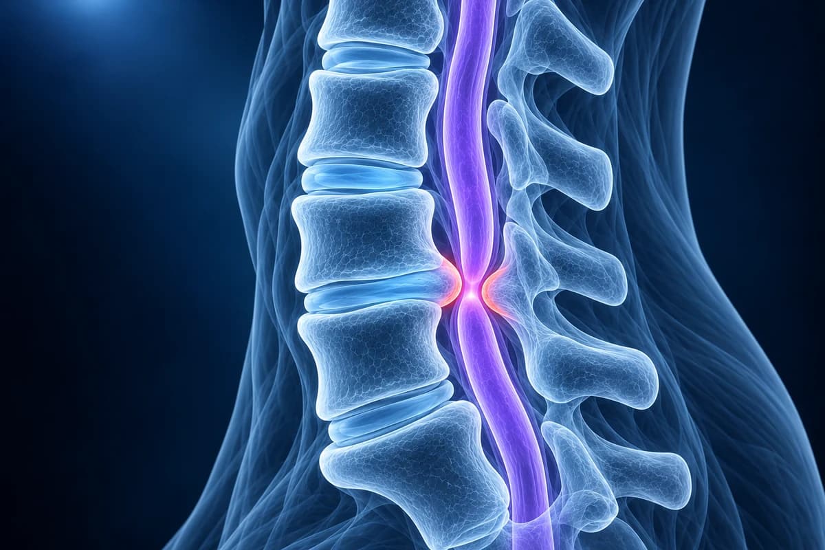
What is degenerative cervical myelopathy?
The spinal cord runs through a bony tunnel in the neck. With age, several changes can make that tunnel smaller at once: discs bulge backward and form a disc osteophyte complex, the ligament behind the canal (ligamentum flavum) thickens and buckles inward, the facet joints enlarge, and one vertebra may slip slightly on another. In some people the ligament along the back of the vertebral bodies turns to bone, a condition called OPLL. People born with a narrower canal reach the point of compression sooner.
The cord is compressed repeatedly as the neck bends and straightens, and its blood supply is reduced. Over time this leads to the symptoms above. Degenerative cervical myelopathy is the most common cause of spinal cord dysfunction in adults, yet it is often recognised late, because early signs such as clumsy fingers or slightly worse balance are easy to put down to age, arthritis or carpal tunnel syndrome.
Myelopathy is different from cervical radiculopathy, where a single nerve root is pinched and the symptoms run down one arm. The two can coexist: a cervical disc herniation or advanced cervical spondylosis can press on both the cord and a root at the same level.
What does "T2 hyperintense cord signal" mean on a neck MRI?
- "T2 hyperintense signal within the cord" or "intramedullary signal change"
- A brighter area inside the cord on T2-weighted images at the level of compression. It reflects swelling, inflammation or early damage in the cord tissue. It supports the diagnosis of myelopathy and is one of the findings surgeons weigh most heavily.
- "T1 hypointense signal" or "myelomalacia"
- A dark area on T1 images at the same spot, or softening of the cord. When a bright T2 change is matched by a dark T1 change, it usually points to longer-standing, less reversible injury, and is associated with a less complete recovery after surgery.
- "Effacement of the CSF" or "effacement of the thecal sac"
- The fluid that normally surrounds the cord has been pushed out of the way at that level. It means the canal is tight, but on its own it does not mean the cord is compressed.
- "Cord flattening", "cord deformity" or "cord compression"
- The cord has lost its normal oval shape and is pressed flat, often from the front by a disc and from behind by thickened ligament. This confirms anatomical compression; whether it causes myelopathy depends on your symptoms and examination.
- "Congenitally narrow canal" or "developmental canal stenosis"
- The bony tunnel was smaller than average from the start. A front-to-back canal diameter below about 13 mm is commonly described as relative stenosis and below about 10 mm as absolute stenosis, which leaves less room for later wear.
For a deeper look at these signs, read cervical myelopathy warning signs on MRI and cervical MRI report phrases explained.
Which scan is used to diagnose cervical myelopathy?
MRI of the cervical spine without contrast is the main test. The ACR Appropriateness Criteria for myelopathy rate it as usually appropriate because it is the only routine study that shows the cord itself, its shape and any signal change, along with the discs and ligaments pressing on it. Contrast is added when infection, tumour or inflammation is a concern rather than simple wear.
CT is better at showing bone and calcified ligament, so it is often added before surgery, especially when OPLL is suspected. CT myelography, where contrast dye is placed in the spinal fluid before the CT, is used when MRI is not possible, for example with some pacemakers, or when metal from earlier surgery blurs the MRI. Upright X-rays, sometimes with flexion-extension views, show overall alignment and whether a vertebra slips with movement. Nerve studies and blood tests are sometimes used to rule out conditions that mimic myelopathy.
What we look for on cervical myelopathy imaging
- Levels where the cord is flattened or deformed, and how many levels are involved
- T2 hyperintense signal inside the cord and any matching T1 hypointense change
- The source of compression: disc osteophyte complex, buckled ligamentum flavum, facet overgrowth or OPLL
- Front-to-back canal diameter and signs of a congenitally narrow canal
- Overall sagittal alignment, including loss of lordosis or kyphosis, and any slip between vertebrae
- Features that suggest a non-degenerative cause, such as a cord lesion away from any compression
Is cord compression on MRI always myelopathy?
No. Some people, particularly older adults, have cord flattening on MRI without any symptoms or abnormal signs. This is sometimes called asymptomatic cord compression. It matters because it raises the chance of developing myelopathy later, and a fall or neck injury can cause more damage than expected. The 2017 AO Spine clinical practice guideline does not recommend preventive surgery for most people with compression but no myelopathy. Instead, it advises explaining the warning symptoms, following them up, and considering surgery if symptoms develop.
Conversely, myelopathy symptoms can come from other causes, such as vitamin B12 deficiency, multiple sclerosis, motor neurone disease or peripheral neuropathy. When the MRI does not show compression at a level that fits, those are considered.
Does cervical myelopathy always need surgery?
Not always, but often. Degenerative cervical myelopathy usually progresses gradually or in steps, and surgery aims to stop further damage rather than to reverse all existing symptoms. Severity is commonly scored with the modified Japanese Orthopaedic Association (mJOA) scale. The 2017 AO Spine clinical practice guideline recommends surgery for moderate and severe myelopathy, and says that for mild myelopathy either surgery or a supervised trial of structured rehabilitation can be offered, with surgery if symptoms worsen.
Operations decompress the cord from the front (anterior cervical discectomy or corpectomy with fusion), from behind (laminoplasty, or laminectomy with fusion), or both. The choice depends on how many levels are involved, where the compression comes from, and the curve of the neck. Earlier treatment is generally linked with better recovery, which is why progressive hand or walking problems deserve attention. Your spine surgeon will explain which approach suits your images. If you already had surgery, see cervical MRI after fusion.
Compression of the lower spinal canal in the lumbar region causes leg symptoms with walking, not cord damage, because the cord ends higher up. It is a different condition from cervical myelopathy.
Can I view my own cervical MRI for cord compression?
You can open your DICOM files privately in the free cervical MRI viewer and compare the sagittal T2 images with the level your report names. A CT can be opened in the free CT viewer. For a plain-language explanation of your images and report, upload them for an AI review. It explains findings; it is not a diagnosis, and new or worsening symptoms should always go to a clinician first.
Frequently Asked Questions
What are the early signs of cervical myelopathy?
Early signs are often subtle: fingers that feel thick or numb in both hands, more difficulty with fine tasks such as buttons or typing, slightly worse handwriting, and a feeling of being less steady on uneven ground. Neck pain may be mild or absent. Anyone with these changes should see a clinician, who can check reflexes and gait.
Is T2 signal change in the spinal cord permanent?
Not always. Some T2 signal change reflects swelling that can fade after the pressure is removed, while change that is also dark on T1 images tends to reflect more lasting damage. Follow-up MRI after treatment helps show how the cord has responded.
How is myelopathy different from a pinched nerve?
A pinched nerve (radiculopathy) affects one nerve root and usually causes pain or numbness down one arm. Myelopathy affects the spinal cord, so symptoms are often in both hands and can involve walking, balance and bladder control. Myelopathy is generally treated with more urgency.
Can physiotherapy fix cervical myelopathy?
Exercise cannot remove the bone or disc pressing on the cord. For mild myelopathy, a supervised rehabilitation programme with close monitoring is a reasonable option under guideline recommendations, but moderate or worsening myelopathy is usually treated surgically. Avoid neck manipulation unless your spine specialist has approved it.
Does a normal neck X-ray rule out myelopathy?
No. X-rays show bone and alignment but not the spinal cord. If myelopathy is suspected, MRI is the test that shows whether and where the cord is compressed.
Sources
- Expert Panel on Neurological Imaging; Agarwal V, Shah LM, Parsons MS, et al. ACR Appropriateness Criteria Myelopathy: 2021 Update. J Am Coll Radiol. 2021;18(5S):S73-S82.
- Fehlings MG, Tetreault LA, Riew KD, et al. A Clinical Practice Guideline for the Management of Patients With Degenerative Cervical Myelopathy: Recommendations for Patients With Mild, Moderate, and Severe Disease and Nonmyelopathic Patients With Evidence of Cord Compression. Global Spine J. 2017;7(3 Suppl):70S-83S.
- Davies BM, Mowforth OD, Smith EK, Kotter MR. Degenerative cervical myelopathy. BMJ. 2018;360:k186.
- Margetis K, Donnally III CJ. Cervical Myelopathy. StatPearls. Treasure Island (FL): StatPearls Publishing.
- American Academy of Orthopaedic Surgeons. Cervical Spinal Cord Compression. OrthoInfo.
Related Articles
What an MRI can and cannot show after an anterior cervical fusion (ACDF): metal artifact, adjacent segment disease, residual compression and cord signal, when CT or X-ray answers better, and which symptoms need urgent care.
When a cervical MRI needs gadolinium (infection, tumour, scar after surgery, MS) and when it does not, plus kidney, retention, allergy and pregnancy safety.
What sets the price of a cervical spine MRI: hospital or imaging centre, contrast (CPT 72141 vs 72156), 1.5T or 3T, insurance, prior authorisation and cash pay.
Do not delay urgent medical care to use this tool.
After you get care, understand your imaging
MRI, X-ray or CT from a CD, folder or ZIP. A PDF report or photos of the films work too.
Free to open and preview, no account needed. An optional AI report from $10 explains your images in plain language, with questions for your doctor.
Your raw files stay on this device. Only rendered images and minimal context are sent when you request an AI report.
Medical Disclaimer: This page is for informational and educational purposes only. It does not constitute medical advice, diagnosis, or treatment. AI-generated analysis may contain errors. Always consult a qualified healthcare professional for medical decisions. Full Disclaimer