
Cervical Spine X-ray Explained: Views, Alignment Lines and Report Terms
What a neck X-ray can and cannot show: the standard views, the four alignment lines, disc space narrowing, bone spurs and the Torg-Pavlov ratio, and when CT or MRI is needed instead.
Key Takeaways
- A neck X-ray shows bones, alignment and disc height, not discs, nerves or the spinal cord
- A good lateral view must reach the C7-T1 junction; four smooth lines should run without steps
- Disc space narrowing and osteophytes are common age changes, most often at C5-C6 and C6-C7
- After significant trauma, adults who need imaging get CT rather than plain X-ray
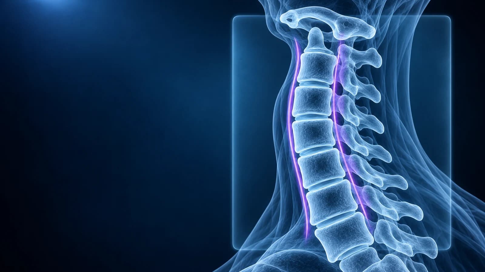
A cervical spine X-ray shows bones, alignment and the height of the disc spaces, but not the discs, nerves or spinal cord themselves. It answers questions like “is the neck lined up?”, “is a bone broken or slipped?” and “how much wear-and-tear (spondylosis) is there?”. It cannot tell you whether a disc is pressing on a nerve; that needs MRI.
Neck X-rays are still common: after a minor injury when a clinician wants a quick check, for long-standing neck pain, to follow a fusion, and to look at movement on bending views. This guide explains each view, the lines a reader traces and the phrases your report is likely to contain. It is educational and does not replace the clinician who reviewed your images.
Which views are in a cervical spine X-ray series?
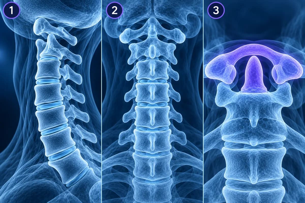
- Lateral (side) view: the most informative single image. It shows alignment, the height of each vertebra and disc space, bone spurs and the soft tissue in front of the spine.
- AP (front-to-back) view: the lower cervical bodies, the spinous processes in a vertical row and the uncovertebral joints along the sides of each disc.
- Open-mouth (odontoid) view: taken through the open mouth to show C1 sitting on C2 and the dens, which the teeth and jaw otherwise hide.
- Oblique views: angled shots that look straight into the nerve exits, so bony narrowing of a foramen can be seen.
- Swimmer’s view: one arm raised above the head to see past the shoulders when the C7-T1 junction is hidden on the plain lateral.
- Flexion and extension views: side views with the head bent forward and back, used to look for abnormal movement between vertebrae. They have their own guide in cervical flexion-extension X-rays.
What are the alignment lines on a lateral neck X-ray?
Four gently curved lines should run smoothly from C2 to C7. The anterior vertebral line follows the front edges of the vertebral bodies. The posterior vertebral line follows their back edges and marks the front wall of the spinal canal. The spinolaminar line joins the points where each lamina meets its spinous process and marks the back wall of the canal. The posterior spinous line connects the tips of the spinous processes. A step in the first two lines means one vertebra has slipped on another (listhesis); a widened gap between two spinous processes can mean a torn ligament.
The overall shape is reported too. A normal neck has a gentle forward curve called lordosis. “Straightening of the cervical lordosis” is very common, often from muscle spasm or simply the way the head was held, and is explained in what straightening of the cervical lordosis means. “Reversal” or “kyphosis” means the curve bends the other way, which matters more when it is focal at one level or appears after an injury.
What do disc space narrowing and osteophytes mean on a neck X-ray?
Discs do not show on X-ray, but the gap they leave between two vertebrae does. When that gap is lower than at the levels above and below, the report says “disc space narrowing” or “loss of disc height”, most often at C5-C6 and C6-C7. Bony lips growing from the edges of the bodies are “osteophytes”; those at the back edge can narrow the canal, and those from the uncovertebral joints can narrow the nerve exits. A hard, sclerotic (whiter) endplate is part of the same process. Together these findings are called cervical spondylosis, and they are extremely common in adults with and without neck pain.
Two patterns deserve a separate look. Thick flowing bone along the front of four or more vertebrae, with the disc spaces kept fairly well, suggests DISH (diffuse idiopathic skeletal hyperostosis). A dense stripe of bone behind the vertebral bodies, inside the canal, can be ossification of the posterior longitudinal ligament (OPLL), which is often confirmed on CT.
Can an X-ray show if the spinal canal is narrow?
Only indirectly. One classic measurement is the Torg-Pavlov ratio: the depth of the spinal canal divided by the depth of the vertebral body on the lateral view, at the same level. Because both are measured on the same film, magnification cancels out. In the original 1987 study a ratio below 0.8 suggested a developmentally narrow canal. The ratio is a screening clue, not a diagnosis, since large vertebral bodies can make it low in people with normal canals. If a narrow canal matters, MRI shows the space around the cord directly.
Is an X-ray enough after a neck injury?
Many people with a minor neck injury need no imaging at all. Two validated decision tools help clinicians decide. The NEXUS criteria say imaging can be skipped when there is no midline neck tenderness, no focal neurological deficit, normal alertness, no intoxication and no painful distracting injury. The Canadian C-spine rule treats age 65 or over, a dangerous mechanism or tingling in the limbs as high risk, and in low-risk patients checks whether the person can actively turn the head 45 degrees to each side.
When adults do need imaging after trauma, the American College of Radiology recommends CT rather than plain X-ray, because CT finds fractures that radiographs miss, especially at C1-C2 and the cervicothoracic junction. MRI is added when the cord, ligaments or neurological symptoms need assessing. For details see cervical spine fracture and whiplash injury.
What does a neck X-ray show at C1 and C2?
On the lateral view, the gap between the front arch of C1 and the dens is the atlanto-dental interval. It should be only a few millimetres and should not widen when the head bends forward. A widened gap suggests the ligament holding the dens in place is lax or torn, which is the core finding of atlantoaxial instability, seen for example in rheumatoid arthritis and Down syndrome. On the open-mouth view, the side edges of C1 should sit over those of C2, and a dark line across the base of the dens can be a fracture or a normal developmental gap, which CT can separate.
When is a neck X-ray not enough?
For neck pain without warning signs, guidelines start with radiographs or with no imaging at all. Arm pain, numbness or weakness that persists points toward MRI, because X-ray cannot show a disc pressing on a nerve root or the cord. Our article do you need an MRI for neck pain explains the usual pathway. Seek same-day care, regardless of a normal X-ray, for neck pain after a significant fall or crash, worsening arm weakness, clumsy hands, unsteady walking, bladder or bowel changes, fever with neck pain, or neck pain with a history of cancer.
Can I look at my own neck X-ray?
Yes. Open the DICOM files in the free X-ray viewer in your browser; the original files stay on your device. Start with the lateral view, adjust brightness until the back edges of the bodies are clear, and trace the lines described above. If your clinician has also ordered an MRI, our guide on how to read a cervical spine MRI picks up where the X-ray stops.
Frequently Asked Questions
Can a neck X-ray show a herniated disc?
No. It can show a narrowed disc space or spurs that suggest wear, but the disc material and any nerve compression need MRI. See cervical disc herniation.
Why was I asked to open my mouth for a neck X-ray?
The open-mouth view lets the beam pass between the teeth to show C1, C2 and the dens, which the jaw otherwise covers. It is part of the standard trauma series.
Is “mild degenerative change” on a neck X-ray serious?
Usually not on its own. Mild narrowing and small spurs are expected with age and correlate poorly with pain. Your symptoms and examination decide whether anything else is needed.
How much radiation is a neck X-ray?
A cervical series uses a low dose, much less than a neck CT. The bigger reason not to repeat X-rays is that they rarely change the plan for ordinary neck pain.
Can AI read my neck X-ray?
AI can explain report terms and point out the structures they describe, which helps you prepare questions. It does not replace the radiologist or the clinician who examined your neck.
Sources
- Expert Panel on Neurological Imaging and Musculoskeletal Imaging; Beckmann NM, West OC, Nunez D Jr, et al. ACR Appropriateness Criteria Suspected Spine Trauma. J Am Coll Radiol. 2019;16(5S):S264-S285.
- Stiell IG, Wells GA, Vandemheen KL, et al. The Canadian C-spine rule for radiography in alert and stable trauma patients. JAMA. 2001;286(15):1841-1848.
- Hoffman JR, Mower WR, Wolfson AB, et al. Validity of a set of clinical criteria to rule out injury to the cervical spine in patients with blunt trauma. N Engl J Med. 2000;343(2):94-99.
- Pavlov H, Torg JS, Robie B, Jahre C. Cervical spinal stenosis: determination with vertebral body ratio method. Radiology. 1987;164(3):771-775.
- Expert Panel on Neurological Imaging; McDonald MA, Kirsch CFE, Amin BY, et al. ACR Appropriateness Criteria Cervical Neck Pain or Cervical Radiculopathy. J Am Coll Radiol. 2019;16(5S):S57-S76.
Related Articles
Why flexion and extension neck X-rays are ordered, how the test is done safely, which measurements suggest instability, why they are not the first test after an injury, and what the report wording means.
What "straightening" or "loss of the normal cervical lordosis" means on a neck X-ray or MRI, why it is common in people without pain, when the curve actually matters, and which findings deserve more attention.
A plain-language guide to your neck MRI: how to count from C2 down to C7, which slices show the cord and the nerve exits, and which artifacts make a cervical scan look worse than it is.
Related Conditions
Understand your imaging before your next appointment
MRI, X-ray or CT from a CD, folder or ZIP. A PDF report or photos of the films work too.
Free to open and preview, no account needed. An optional AI report from $10 explains your images in plain language, with questions for your doctor.
Your raw files stay on this device. Only rendered images and minimal context are sent when you request an AI report.
Medical Disclaimer: This page is for informational and educational purposes only. It does not constitute medical advice, diagnosis, or treatment. AI-generated analysis may contain errors. Always consult a qualified healthcare professional for medical decisions. Full Disclaimer