
Cervical Spondylosis: What Neck Wear-and-Tear Looks Like on Your Report
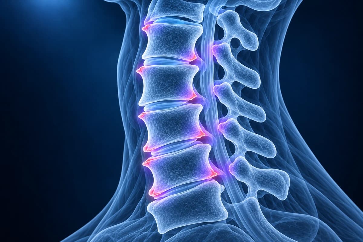
Cervical spondylosis is the umbrella term radiologists use for age-related wear of the neck: thinner discs, bony spurs (osteophytes), enlarged uncovertebral and facet joints. It is extremely common and often painless. It matters mainly when it narrows the openings for nerves (foraminal stenosis) or the canal around the spinal cord (canal stenosis), which a report states separately.
Common Symptoms
- Aching, stiff neck that is often worst in the morning or after holding the head in one position
- Grinding or crunching sensations when turning the head
- Pain spreading to the shoulder blades or the back of the head
- Gradual loss of the ability to look over the shoulder, for example when reversing a car
- Arm pain, tingling or weakness when a narrowed foramen irritates a nerve root
- Clumsy hands or unsteady walking when spurs and discs narrow the canal around the cord
When does neck wear-and-tear need urgent attention?
Spondylosis itself is not an emergency, but see a doctor promptly if you notice progressive weakness or numbness in an arm or hand, clumsy fingers (trouble with buttons, writing or dropping things), a change in your walking or balance, or electric shocks down the spine when bending the neck: these can mean the cord is being compressed. Go to an emergency department the same day for new bladder or bowel problems, rapidly worsening weakness, neck pain after a fall or crash, or neck pain with fever, unexplained weight loss, a history of cancer or a weakened immune system. These symptoms are not explained by ordinary wear.
What does "cervical spondylosis" mean on my report?
The word comes from the Greek for vertebra and simply means degenerative change in the spine. When a report says "multilevel cervical spondylosis" or "degenerative changes most pronounced at C5–C6", it is summarising a cluster of findings rather than naming a single disease. Those findings start with the disc: as it dries out, it loses height, bulges slightly and loads the bone edges differently. The bone responds by growing spurs. The small joints at the side and back of each segment then thicken, and the ligament lining the back of the canal can buckle inward.
Because these changes appear in most adults with age, they show up on scans of people with no neck pain at all. Large reviews of imaging in people without symptoms, and comparisons of neck pain patients with pain-free controls, have found that most degenerative findings are only weakly linked to pain. A spondylosis report is therefore a description, not a diagnosis of why your neck hurts.
Which report phrases describe cervical spondylosis?
- "Uncovertebral hypertrophy" or "uncovertebral joint arthropathy"
- The uncovertebral joints (joints of Luschka) are small ridges at the side edges of the C3 to C7 vertebral bodies that exist only in the neck. When they enlarge they grow toward the foramen, the exit tunnel for the nerve root, and are a leading cause of nerve narrowing in the neck.
- "Marginal osteophytes" or "anterior osteophytes"
- Bony spurs at the corners of the vertebral bodies. Those at the front rarely matter unless very large; posterior ones point toward the canal and the cord.
- "Disc height loss" or "disc space narrowing"
- The disc has thinned. It often comes with "disc desiccation", meaning the disc looks darker on T2 MRI because it holds less water.
- "Disc osteophyte complex"
- A broad ridge at the back of the disc where bulging disc material and bony spurs merge and cannot be told apart on MRI. The report then says whether it touches or flattens the thecal sac or cord. Our disc osteophyte complex explainer goes further.
- "Facet arthropathy" and "ligamentum flavum thickening"
- Arthritis of the small paired joints at the back, and a thicker ligament lining the back of the canal. Both can narrow the canal and foramina from behind.
- "Degenerative anterolisthesis"
- One vertebra has slipped a few millimetres forward on the one below because the facet joints are worn. In the neck this is most often seen at C3–C4 and C4–C5, above stiffer lower levels.
For the full glossary, see the cervical MRI report phrases guide.
What is the difference between spondylosis and stenosis?
Spondylosis is the process; stenosis is one possible consequence. A report can describe advanced spondylosis with widely open foramina and a roomy canal, which usually means no nerve is being squeezed. Stenosis means a space has become narrow, and radiologists grade it as mild, moderate or severe for each level and side.
- "Foraminal narrowing" or "foraminal stenosis" means the side exit for a nerve root is tighter, typically from uncovertebral spurs. It can cause arm pain in a nerve root pattern, described on our cervical foraminal stenosis and cervical radiculopathy pages.
- "Central canal stenosis" means the main canal is narrowed, from disc osteophyte complexes in front and a buckled ligamentum flavum behind. When the cord is flattened, especially with a bright spot on T2 images, the concern is cervical myelopathy.
- "Effacement of the ventral CSF" means the fluid in front of the cord is pushed aside, but the cord itself is not yet deformed. It is a milder finding than "cord compression".
Lumbar and whole-spine stenosis follow similar principles but produce different symptoms; this page stays with the neck.
What we look for on spondylosis imaging
- Disc height loss and desiccation at each level from C2–C3 to C7–T1
- Uncovertebral and facet joint hypertrophy and the side they narrow
- Disc osteophyte complexes and their contact with the thecal sac or cord
- Foraminal narrowing graded per level and side on oblique or axial images
- Central canal narrowing, cord flattening and any T2 hyperintense cord signal
- Alignment: loss of lordosis, kyphosis and degenerative slips
Do you need an X-ray or MRI for cervical spondylosis?
For chronic neck pain associated with spondylosis and radiculopathy, without trauma or red flags, ACR criteria rate MRI without contrast as usually appropriate. Without radiculopathy or red flags, cervical radiographs or non-contrast MRI may be appropriate.
For neck pain without arm symptoms or red flags, imaging is usually not needed at first, because it rarely changes treatment and almost always shows some spondylosis. When imaging is done, plain X-rays show disc height loss, spurs, alignment and, on oblique views, the bony outline of the foramina; see the cervical spine X-ray explainer. MRI is the test when there is persistent arm pain, neurological signs, or symptoms of cord compression, because it shows the nerves, cord and discs directly. CT shows bony spurs and ossified ligaments best and is used for surgical planning or when MRI is not possible. Our article on whether you need an MRI for neck pain walks through when imaging actually helps.
Some findings look like spondylosis but are separate conditions. Flowing bone bridging the front of four or more vertebrae with preserved discs suggests diffuse idiopathic skeletal hyperostosis (DISH), and a calcified band behind the vertebral bodies suggests ossification of the posterior longitudinal ligament (OPLL).
How is cervical spondylosis treated?
Most people with neck pain from spondylosis are treated without surgery: staying active, neck and upper back exercise guided by a physiotherapist, posture and workstation changes, and short courses of pain relief. Flare-ups often settle over weeks. Injections may be considered for selected nerve root or facet pain. Surgery, such as anterior cervical discectomy and fusion, disc replacement or a posterior decompression, is reserved for nerve root compression with persistent pain or weakness, or for cord compression. Spondylosis seen on a scan is not by itself a reason for an operation. Your clinician combines the images with your examination before recommending any of these. If you have already had a fusion, our guide to cervical MRI after fusion explains what changes at the levels next to it.
Can I look through my own neck MRI or X-ray?
Yes. Open your study privately in the free neck MRI viewer or, for plain films, the free X-ray viewer; the files stay on your device. If you want each level's wording translated into plain language, upload the images for an AI review. It explains findings; it does not diagnose and does not replace your doctor or radiologist.
Frequently Asked Questions
Is cervical spondylosis the same as arthritis of the neck?
Broadly, yes. Spondylosis covers wear of the discs and the small joints, and the facet and uncovertebral joint changes are a form of osteoarthritis. It is different from inflammatory arthritis such as rheumatoid arthritis, which affects the neck in other ways.
Does cervical spondylosis get worse over time?
The imaging changes usually progress slowly with age, but symptoms do not track them closely. Many people have long pain-free periods with occasional flare-ups. A small number develop nerve root or cord compression, which is why new arm weakness, clumsy hands or walking changes should be reported.
What does "moderate uncovertebral hypertrophy with foraminal narrowing" mean?
The bony ridges at the side of the disc have enlarged and partly narrowed the exit for a nerve root at that level. Whether it matters depends on whether you have arm symptoms in that nerve's distribution. Many people with this wording have no arm symptoms at all.
Can cervical spondylosis cause headaches or dizziness?
Upper neck joint changes can refer pain to the back of the head, a pattern described on our cervicogenic headache page. Dizziness has many causes, most outside the neck, so it should be assessed by a clinician rather than attributed to a scan finding.
Sources
- Theodore N. Degenerative cervical spondylosis. N Engl J Med. 2020;383(2):159-168.
- Margetis K, Tadi P. Cervical Spondylosis. StatPearls. Treasure Island (FL): StatPearls Publishing.
- Brinjikji W, Luetmer PH, Comstock B, et al. Systematic literature review of imaging features of spinal degeneration in asymptomatic populations. AJNR Am J Neuroradiol. 2015;36(4):811-816.
- Farrell SF, Smith AD, Hancock MJ, et al. Cervical spine findings on MRI in people with neck pain compared with pain-free controls: a systematic review and meta-analysis. J Magn Reson Imaging. 2019;49(6):1638-1654.
Related Articles
What "straightening" or "loss of the normal cervical lordosis" means on a neck X-ray or MRI, why it is common in people without pain, when the curve actually matters, and which findings deserve more attention.
What a neck X-ray can and cannot show: the standard views, the four alignment lines, disc space narrowing, bone spurs and the Torg-Pavlov ratio, and when CT or MRI is needed instead.
What your neck MRI report means: uncovertebral hypertrophy, foraminal narrowing grades, effacement of the CSF, cord flattening, T2 hyperintense cord signal, Modic changes and more, each explained in one or two plain sentences.
Understand your imaging before your next appointment
MRI, X-ray or CT from a CD, folder or ZIP. A PDF report or photos of the films work too.
Free to open and preview, no account needed. An optional AI report from $10 explains your images in plain language, with questions for your doctor.
Your raw files stay on this device. Only rendered images and minimal context are sent when you request an AI report.
Medical Disclaimer: This page is for informational and educational purposes only. It does not constitute medical advice, diagnosis, or treatment. AI-generated analysis may contain errors. Always consult a qualified healthcare professional for medical decisions. Full Disclaimer