
Whiplash MRI: What It Shows, What It Misses and When You Need One
Why most whiplash MRIs look normal, which injuries MRI can find (ligament tears, bone bruises, disc injury, cord contusion), what report phrases like prevertebral edema mean, and when imaging is needed after a crash.
Key Takeaways
- Most whiplash MRIs are normal because muscle and joint strain is too subtle to see
- Numbness, weakness, midline bone pain, a dangerous mechanism or age 65 and over need emergency assessment
- After a crash, CT usually comes first for bone; MRI is added for nerve symptoms or suspected ligament injury
- MRI can show ligament tears, bone bruises, facet joint fluid, traumatic disc injury and cord contusion when they are present
- Bright signal in the alar or transverse ligaments alone was not linked to whiplash in a meta-analysis
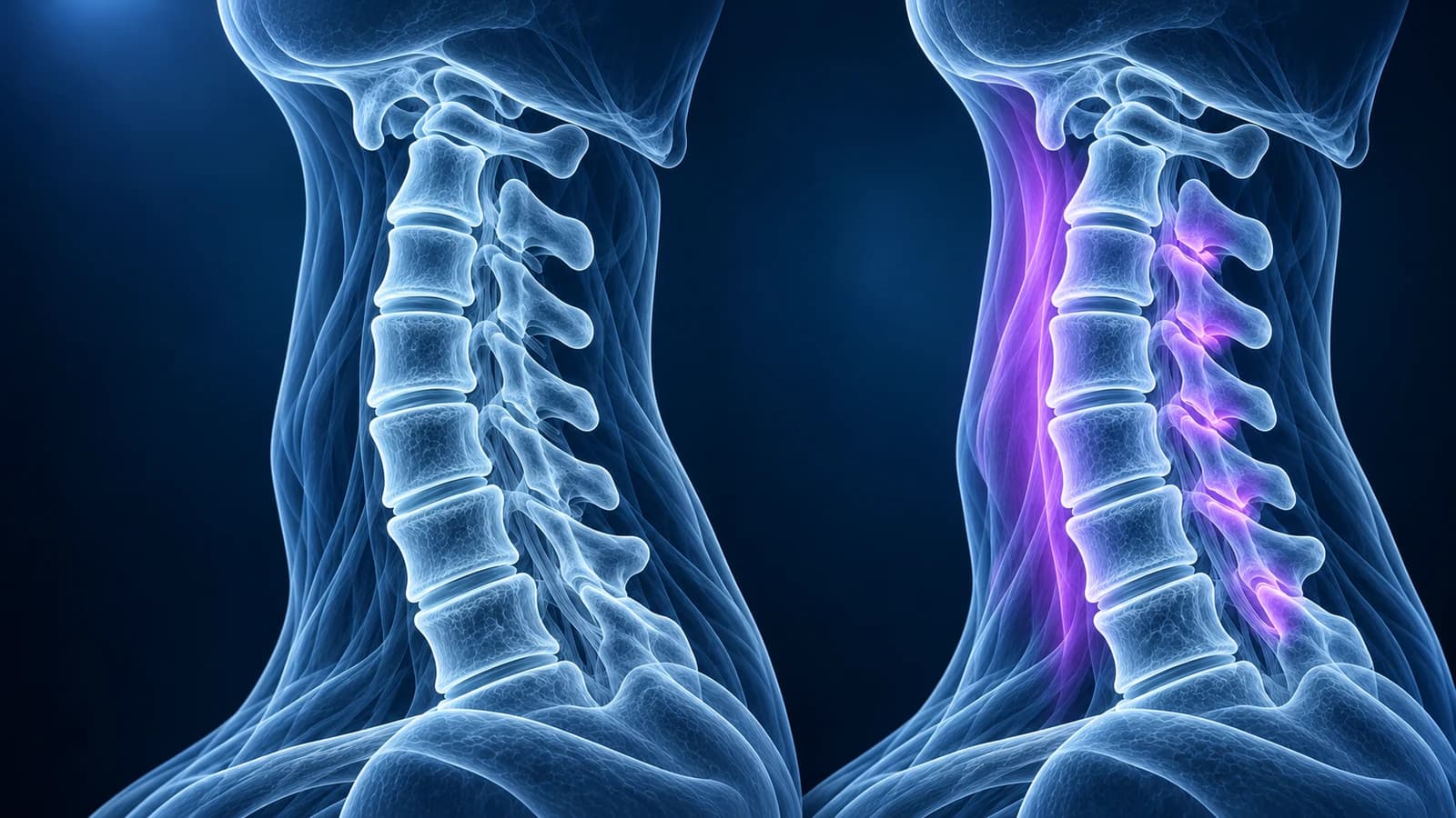
For most people with whiplash, the MRI is normal or shows only age-related wear that was there before the crash. Whiplash is mainly a strain of muscles, small joints and ligaments, and those micro-injuries are usually too small for MRI to see. MRI is used when there are nerve or spinal cord symptoms, or when a ligament tear, fracture or disc injury needs to be ruled out.
That can feel frustrating. Pain after a collision is real whether or not a scan can photograph its cause, and a normal MRI is not a judgement that nothing happened. It means the serious structural injuries the scan is designed to catch were not found. This guide explains what MRI can and cannot see after whiplash, how reports word those findings, and when a scan is the right next step. It is educational, not a diagnosis of your neck.
When is neck pain after a crash an emergency?
Call emergency services or go to an emergency department, keeping the neck as still as possible, if neck pain after a collision or fall comes with any of these. Decisions about imaging are made there, not at home.
- Numbness, tingling or weakness in the arms or legs, or a feeling of electric shocks down the spine
- Pain directly over the bones at the back of the neck, or inability to turn the head to either side
- A high-speed or rollover collision, ejection, a fall from height or a diving injury
- Age 65 or older, or a condition that stiffens the spine such as ankylosing spondylitis
- Loss of consciousness, confusion, severe headache, trouble swallowing or a change in voice
- Difficulty walking, or new problems controlling the bladder or bowel
Does whiplash show up on an MRI?
In the usual case, no. During a rear-end impact the torso is pushed forward while the head lags, so the neck briefly takes an S-shaped curve before it whips into extension and then flexion. The load falls on the facet joint capsules, the deep neck muscles and the ligaments that join one vertebra to the next. Stretching those tissues causes pain, stiffness and spasm without tearing them across, and stretched tissue looks the same as healthy tissue on MRI.
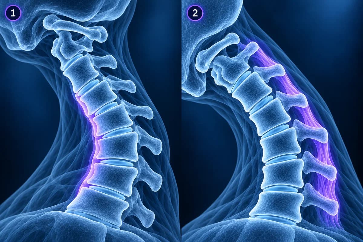
The Quebec Task Force on Whiplash-Associated Disorders grades the condition by examination rather than by scan. Grade I is neck pain, stiffness or tenderness only. Grade II adds physical signs such as reduced movement and point tenderness. Grade III adds neurological signs such as weakness or reduced reflexes. Grade IV means a fracture or dislocation. MRI changes management mainly at grades III and IV, which is why it is not routine for grades I and II.
What can a whiplash MRI find?
When the injury is more than a strain, MRI is the test that shows soft tissue, marrow and the spinal cord. Its fluid-sensitive images, especially STIR, make fresh injury bright against a dark background, which is how acute damage is separated from old wear. The findings below are the ones radiologists look for after a neck injury.
- Ligament injury: bright fluid or a visible gap in the anterior longitudinal ligament at the front of the spine, or in the interspinous ligaments between the bony points at the back
- Bone bruise: patchy marrow edema in a vertebral body or facet without a fracture line, a sign of impact rather than a break
- Facet joint effusion or capsular edema: fluid in or around the small joints at the back of the neck, which took much of the whiplash load
- Traumatic disc injury: a disc tearing from the bone edge or a fresh herniation that is new compared with the wear at other levels
- Spinal cord contusion or epidural hematoma: rare but serious findings in people with neurological symptoms
Fractures themselves are better shown by CT, and an injury that makes the upper neck unstable is assessed with both. Our pages on cervical spine fracture and atlantoaxial instability describe those injuries in more detail.
What do whiplash MRI report phrases mean?
- “Prevertebral soft tissue edema” — fluid in the soft tissue in front of the vertebrae, a marker that the front of the spine was stretched hard, often prompting a closer look at the ligaments
- “Interspinous ligament edema” — bright signal between the spinous processes at the back, suggesting a sprain or partial tear of the posterior ligaments
- “No acute osseous or ligamentous injury” — no fresh fracture, bone bruise or visible ligament tear; muscle and joint strain are still possible
- “Straightening of the cervical lordosis, likely positional or related to muscle spasm” — the neck looks less curved than usual, common after whiplash and explained in straightening of the cervical lordosis
- “Chronic degenerative changes without acute abnormality” — wear that pre-dates the crash; it can make a strained neck slower to settle but is not itself the injury
- “Cord signal is normal” — no swelling or bruising inside the spinal cord, one of the most reassuring lines after a neck injury
More wording is decoded in cervical MRI report phrases explained.
What about bright signal in the alar or transverse ligaments?
The alar and transverse ligaments hold the top two vertebrae to the skull and to each other, and some clinics promote high-resolution or upright MRI to look for whiplash damage there. A meta-analysis of case-control studies found that bright signal in these ligaments was not linked to whiplash-associated disorders: it appeared about as often in people without whiplash. The authors concluded that MRI of these ligaments should not be part of the routine work-up. A true disruption with instability is a different matter and is judged on alignment and movement, not on signal alone.
When do you need an MRI after whiplash?
The first decision is about bone, not soft tissue. Emergency clinicians use the Canadian C-spine rule or the NEXUS criteria to decide whether any imaging is needed; for example, an alert adult after a simple rear-end collision with no midline tenderness who can turn the head 45 degrees to each side does not need a scan. When imaging is needed, the ACR guidance for suspected spine trauma favours CT for adults as the first test.
MRI is then added for arm or leg symptoms, weakness, a CT finding that suggests ligament injury, alignment that looks wrong despite no fracture, or pain that stays severe and unexplained. Later on, persistent arm pain in a nerve pattern weeks after the crash is assessed the same way as any cervical radiculopathy. If instability is the question once the acute pain allows movement, flexion-extension X-rays show whether a level shifts when the head bends. For the wider picture, see do you need an MRI for neck pain.
Why does my neck still hurt if the MRI is normal?
Strained facet joint capsules, irritated muscles and a sensitised nervous system all generate pain without leaving a mark on MRI. Most people with whiplash improve over weeks to months, and staying active with guided exercise is generally preferred to collars and prolonged rest. Persistent headache from the upper neck, sometimes after whiplash, is covered in cervicogenic headache, and the condition itself in whiplash injury. A clinician who examines you remains the right person to decide whether further tests are useful.
If you have the DICOM files from the scan, the free MRI viewer and free CT viewer open them in your browser, so you can look at the slices your report refers to without uploading them anywhere.
Frequently Asked Questions
Can an MRI prove a whiplash injury for an insurance claim?
Usually not on its own. A typical whiplash strain leaves no visible change, and degenerative findings on the scan generally pre-date the crash. Clinical records from the time of the injury, the examination and the course of symptoms carry more weight than a normal or age-typical MRI. This is general information, not legal advice.
How soon after a car accident should a neck MRI be done?
If there are neurological symptoms or a suspected ligament injury, MRI is done urgently, ideally within days, because acute edema is easiest to see early and fades over weeks. Without those features, most people do not need an MRI at all, and one taken months later is used to look for a persistent nerve or disc problem rather than the original strain.
Can whiplash cause a herniated disc in the neck?
It can, but it is less common than many people assume. MRI after a crash often shows disc bulges that are equally common in uninjured adults. Signs that point toward a new injury include fresh marrow edema at the same level, a herniation that matches new arm symptoms, and a level that looks different from its neighbours. See cervical disc herniation for more.
Is an upright or dynamic MRI better for whiplash?
Upright and positional MRI can show the neck under load, but they are not part of standard whiplash pathways, and their findings, particularly in the upper-neck ligaments, have not been shown to separate injured from uninjured necks reliably. Standard MRI plus flexion-extension X-rays when instability is suspected remains the usual approach.
Sources
- Spitzer WO, Skovron ML, Salmi LR, et al. Scientific monograph of the Quebec Task Force on Whiplash-Associated Disorders: redefining "whiplash" and its management. Spine (Phila Pa 1976). 1995;20(8 Suppl):1S-73S.
- Li Q, Shen H, Li M. Magnetic resonance imaging signal changes of alar and transverse ligaments not correlated with whiplash-associated disorders: a meta-analysis of case-control studies. Eur Spine J. 2013;22(1):14-20.
- Expert Panel on Neurological Imaging and Musculoskeletal Imaging; Beckmann NM, West OC, et al. ACR Appropriateness Criteria Suspected Spine Trauma. J Am Coll Radiol. 2019;16(5S):S264-S285.
- Stiell IG, Wells GA, Vandemheen KL, et al. The Canadian C-spine rule for radiography in alert and stable trauma patients. JAMA. 2001;286(15):1841-1848.
Related Articles
What "straightening" or "loss of the normal cervical lordosis" means on a neck X-ray or MRI, why it is common in people without pain, when the curve actually matters, and which findings deserve more attention.
Why flexion and extension neck X-rays are ordered, how the test is done safely, which measurements suggest instability, why they are not the first test after an injury, and what the report wording means.
Most neck pain does not need an MRI. Learn the warning signs that do call for imaging, why X-ray or CT comes first after an injury, and how to read the age-related findings a neck MRI almost always shows.
Related Conditions
Understand your imaging before your next appointment
MRI, X-ray or CT from a CD, folder or ZIP. A PDF report or photos of the films work too.
Free to open and preview, no account needed. An optional AI report from $10 explains your images in plain language, with questions for your doctor.
Your raw files stay on this device. Only rendered images and minimal context are sent when you request an AI report.
Medical Disclaimer: This page is for informational and educational purposes only. It does not constitute medical advice, diagnosis, or treatment. AI-generated analysis may contain errors. Always consult a qualified healthcare professional for medical decisions. Full Disclaimer