
Whiplash Injury: Normal MRI, Real Pain and the Findings That Do Matter
Whiplash is a strain of the soft tissues of the neck caused by a sudden back-and-forth movement, most often in a rear-end car crash. Most people with whiplash have a normal X-ray and a normal or age-typical MRI, because the injury is usually to muscles, small joints and ligaments at a level scans cannot resolve. A normal scan does not mean the pain is imagined; it means no fracture, instability or nerve compression was found.
Common Symptoms
- Neck pain and stiffness that often starts or worsens several hours to a day after a rear-end collision
- Pain spreading into the shoulders, between the shoulder blades or up to the base of the skull
- Headache that begins at the back of the head, typically worse with neck movement
- Reduced ability to turn or tilt the head, with tender muscles along the back of the neck
- Tiredness, dizziness, poor concentration or disturbed sleep in the first weeks after the crash
- Tingling or aching in an arm, which needs a clinical check for nerve root irritation
When is neck pain after a crash an emergency?
Whiplash is a diagnosis made only after a more serious injury has been ruled out. Call emergency services or go to an emergency department the same day if, after any collision or fall, you have weakness, numbness or electric shocks in the arms or legs, trouble walking or keeping your balance, loss of bladder or bowel control, severe midline neck pain directly over the bones, a feeling that the head is unstable, a severe or worsening headache, double vision, slurred speech, difficulty swallowing, or confusion. Older adults and people with a stiff spine from ankylosing spondylitis or DISH can break the neck in a low-speed crash, so they should be examined even when the impact felt minor. Do not let anyone manipulate your neck until a fracture has been excluded.
If a fracture is a possibility, read our page on cervical spine fractures, which explains the CT pathway used in emergency departments.
What actually happens to the neck in a whiplash injury?
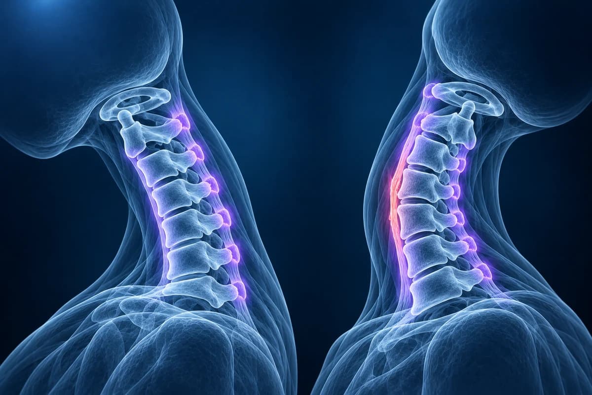
High-speed film studies of rear-end impacts show the torso being shoved forward by the seat while the head stays put for a fraction of a second. For a moment the neck forms an S-shape: the lower segments bend backward while the upper segments bend forward. That unnatural curve compresses the back of the small facet joints and stretches the front of the discs and the anterior longitudinal ligament, before the head snaps forward again. Side impacts and a head that is turned at the moment of the crash load the joints asymmetrically.
The tissues most often blamed for persistent whiplash pain are the facet joint capsules, the neck muscles and the ligaments that hold each segment together. These injuries are microscopic stretches and tears rather than large ruptures, which is why they rarely produce a visible change on imaging. Recovery is usually good: many people improve substantially within weeks, while a minority have pain that lasts months. Early return to normal activity, simple pain relief and graded exercise are the usual first-line approach.
How is whiplash graded?
Clinicians still use the Quebec Task Force classification of whiplash-associated disorders, published in 1995. It grades the injury by examination findings, not by scans:
- Grade 0: no neck complaints and no physical signs.
- Grade I: neck pain, stiffness or tenderness only, with no physical signs on examination.
- Grade II: neck complaints plus musculoskeletal signs such as reduced range of movement and point tenderness.
- Grade III: neck complaints plus neurological signs such as reduced reflexes, weakness or sensory loss.
- Grade IV: neck complaints with a fracture or dislocation.
Grades I and II make up most cases and are soft-tissue injuries by definition. Grade III points toward an irritated nerve root, which is assessed like cervical radiculopathy. Grade IV is no longer whiplash in the everyday sense; it is a spinal fracture or dislocation.
Do you need an X-ray, CT or MRI after whiplash?
Many people do not need any scan. Emergency clinicians use validated decision rules to decide who needs imaging after neck trauma. The Canadian C-spine rule, for alert and stable adults, recommends imaging for anyone aged 65 or over, anyone with a dangerous mechanism (such as a high-speed or rollover crash, ejection from a vehicle, or a fall from about 1 metre or five stairs) and anyone with tingling in the limbs. Patients without those high-risk factors who have a low-risk feature, such as a simple rear-end collision or the ability to walk around after the crash, and who can actively turn the head 45 degrees to each side, can usually be cleared without imaging.
When imaging is needed in an adult, the ACR Appropriateness Criteria for suspected spine trauma favour CT of the cervical spine over plain X-rays, because CT finds fractures that radiographs miss. MRI is added when there are neurological signs, when CT is normal but a ligament injury is still suspected, or when pain and nerve symptoms persist. For a deeper look at this question, read whether you need an MRI for neck pain.
Why is my whiplash MRI normal when my neck still hurts?
In a prospective study of 100 people scanned within three weeks of a whiplash injury, MRI showed an injury-related abnormality in only one of them, and later research has not found a scan pattern that reliably predicts who will recover. Strained muscle fibres and stretched joint capsules simply do not change MRI signal enough to be seen, and any mild swelling fades within days to weeks.
The opposite problem is just as common: an MRI after a crash often lists disc bulges, small osteophytes or "straightening of the cervical lordosis". These are frequent in people with no neck pain at all and were usually present before the accident. Straightening of the curve in particular is often caused by muscle spasm or simply by lying flat in the scanner; our explainer on straightening of the cervical lordosis covers it. Background wear is described on the cervical spondylosis page. Separating age-related changes from new injury is one of the main jobs of the radiologist and your treating clinician.
What does a positive whiplash finding look like on a report?
When an acute neck injury does show on MRI, the report usually uses wording like the phrases below. Fluid-sensitive sequences such as STIR are the ones that show fresh injury, and timing matters: these signs are most visible in the first days to weeks.
- "Prevertebral soft tissue swelling" or "prevertebral edema"
- Fluid or blood in the thin layer of tissue in front of the vertebrae. It can follow a tear of the front ligament or a hidden fracture, so it prompts a careful look for instability rather than being dismissed.
- "Anterior longitudinal ligament disruption"
- The long ligament running down the front of the vertebral bodies is torn, typically from forced extension. With disc widening at the same level it can mean the segment is unstable.
- "Interspinous ligament edema"
- Bright fluid signal between the spinous processes at the back of the neck, a sign of a flexion strain of the posterior ligament complex. Isolated mild edema is common; edema with widened spinous processes is more significant.
- "Facet joint effusion" or "facet capsular edema"
- Extra fluid in or around one of the small paired joints at the back of the spine. After trauma it may reflect a capsular sprain; in older people a small effusion often accompanies arthritis instead.
- "Vertebral body bone marrow edema"
- Bruising inside a vertebra. It means the bone took a significant load and is a reason to check the CT again for a subtle fracture line.
Our guide to cervical MRI report phrases explains more wording, and what a whiplash MRI can and cannot show compares normal and strained soft tissues side by side.
What we look for on post-whiplash imaging
- Prevertebral soft tissue thickening in front of C2 to C6 compared with expected thickness
- Signal change or discontinuity of the anterior longitudinal ligament with disc space widening
- Interspinous or ligamentum flavum edema and widening between spinous processes
- Facet joint effusion, perched or subluxed facets, or loss of facet joint alignment
- Vertebral body or endplate marrow edema hinting at an occult fracture
- New disc herniation or cord signal change in patients with arm or leg symptoms
What else can explain symptoms after a whiplash injury?
Headache at the back of the skull that persists after a crash may come from the upper neck joints, described on our cervicogenic headache page; a severe, sudden or worsening headache is different and needs urgent assessment for bleeding or arterial injury. Arm pain in a dermatome, with numbness or weakness, can come from a cervical disc herniation that the crash aggravated. In people who already had a narrow canal, an extension injury can bruise the cord without any fracture, which is why clumsy hands or unsteady walking after a crash always need an MRI. Persisting dizziness, memory or concentration problems may also reflect a concussion, which is assessed separately from the neck.
Can I review my own neck scan after a car accident?
If you were given a disc with your images, you can open them privately in the free neck MRI viewer or, for an emergency department scan, the free CT viewer. Files stay on your device. For a plain-language explanation of the report wording and what the images show, upload them for an AI review. It is not a diagnosis and it does not replace the radiologist, your doctor or a medico-legal assessment.
Frequently Asked Questions
Can an MRI prove I have whiplash?
Usually not. Whiplash is a clinical diagnosis based on the history of the crash and the examination. MRI is used to exclude problems that change treatment, such as a ligament rupture, a disc herniation pressing on a nerve, or cord injury. A normal MRI is the expected result in most whiplash cases and is reassuring rather than a sign that nothing happened.
How soon after a crash should a whiplash MRI be done?
Immediate MRI is reserved for people with neurological signs or suspected ligament instability. For ordinary whiplash, guidelines suggest treating symptoms and reassessing; MRI is considered if arm symptoms, weakness or severe pain persist beyond several weeks. If an acute ligament injury is the question, scanning within the first days to two weeks is best because swelling fades after that.
Does a disc bulge on my MRI mean the accident caused it?
Not necessarily. Disc bulges and small osteophytes are common in people of every age who have never had neck pain. Clues that a finding is new include fluid signal (edema) around it, matching nerve symptoms, and comparison with any earlier scans. Your radiologist and treating clinician weigh these together.
How long does whiplash take to heal?
Many people improve markedly within the first few weeks to three months, especially when they stay active. Higher initial pain, early arm symptoms and high distress are associated with slower recovery. Pain that keeps getting worse rather than better, or new nerve symptoms, should be reviewed by a clinician.
Should I wear a soft collar after whiplash?
For uncomplicated whiplash, prolonged collar use is generally discouraged because early, gentle movement is linked to better recovery. A rigid collar is a different matter: it is used when a fracture or instability has not yet been excluded, and should only be removed when a clinician says so.
Sources
- Spitzer WO, Skovron ML, Salmi LR, et al. Scientific monograph of the Quebec Task Force on Whiplash-Associated Disorders: redefining "whiplash" and its management. Spine (Phila Pa 1976). 1995;20(8 Suppl):1S-73S.
- Stiell IG, Wells GA, Vandemheen KL, et al. The Canadian C-spine rule for radiography in alert and stable trauma patients. JAMA. 2001;286(15):1841-1848.
- Ronnen HR, de Korte PJ, Brink PR, et al. Acute whiplash injury: is there a role for MR imaging? A prospective study of 100 patients. Radiology. 1996;201(1):93-96.
- Farrell SF, Smith AD, Hancock MJ, et al. Cervical spine findings on MRI in people with neck pain compared with pain-free controls: a systematic review and meta-analysis. J Magn Reson Imaging. 2019;49(6):1638-1654.
Related Articles
What "straightening" or "loss of the normal cervical lordosis" means on a neck X-ray or MRI, why it is common in people without pain, when the curve actually matters, and which findings deserve more attention.
Why most whiplash MRIs look normal, which injuries MRI can find (ligament tears, bone bruises, disc injury, cord contusion), what report phrases like prevertebral edema mean, and when imaging is needed after a crash.
Why flexion and extension neck X-rays are ordered, how the test is done safely, which measurements suggest instability, why they are not the first test after an injury, and what the report wording means.
Understand your imaging before your next appointment
MRI, X-ray or CT from a CD, folder or ZIP. A PDF report or photos of the films work too.
Free to open and preview, no account needed. An optional AI report from $10 explains your images in plain language, with questions for your doctor.
Your raw files stay on this device. Only rendered images and minimal context are sent when you request an AI report.
Medical Disclaimer: This page is for informational and educational purposes only. It does not constitute medical advice, diagnosis, or treatment. AI-generated analysis may contain errors. Always consult a qualified healthcare professional for medical decisions. Full Disclaimer