
OPLL: Ossification of the Posterior Longitudinal Ligament on CT and MRI
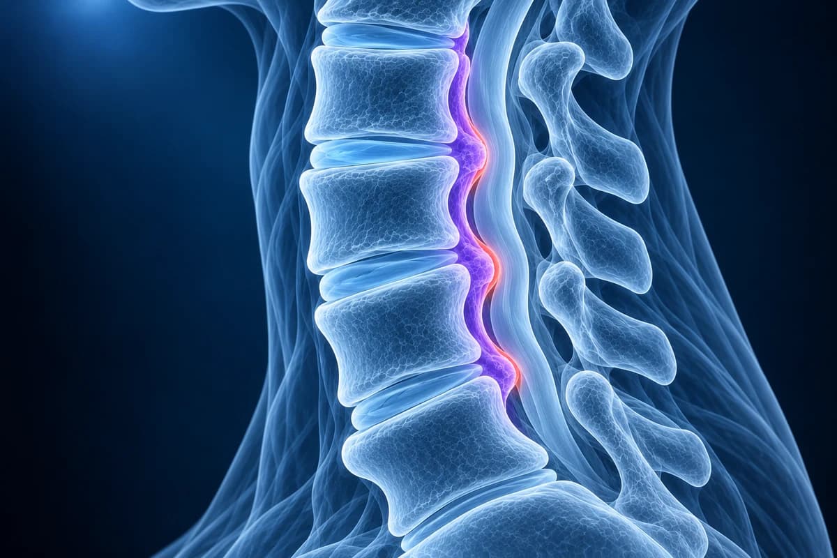
OPLL (ossification of the posterior longitudinal ligament) means the ligament running down the back of the vertebral bodies, directly in front of the spinal cord, has gradually turned to bone. CT shows it best; MRI shows how much it presses on the cord. Many people have no symptoms, but thick OPLL can cause myelopathy and changes how neck surgery is planned.
Common Symptoms
- No symptoms at all in many people, with OPLL first noticed on an X-ray or CT taken for another reason
- Neck stiffness and a gradual loss of the ability to tip the head back
- Numbness or clumsiness in both hands, especially with fine tasks such as buttons or chopsticks
- Unsteady walking or legs that feel stiff and heavy
- Arm pain or tingling when the ossification also narrows a nerve opening
- Sudden arm or leg weakness after a minor fall or whiplash-type jolt in someone with a narrowed canal
When is OPLL an emergency?
A canal already narrowed by OPLL leaves the cord little spare room. After a fall, a car crash or even a minor jolt that bends the neck backward, go to an emergency department straight away if you notice new weakness, numbness or burning in the arms or hands, difficulty walking, or bladder or bowel changes. These can be signs of a cord injury without any fracture. Outside of injury, progressive hand clumsiness or a change in balance needs prompt review by a spine specialist, as described on our cervical myelopathy page.
What is the posterior longitudinal ligament, and why does it ossify?
The posterior longitudinal ligament is a strong band that runs along the back surfaces of the vertebral bodies and discs, from the base of the skull downward. It forms the front wall of the spinal canal. In OPLL, ligament tissue is replaced step by step by mature bone. The process usually starts in the neck, most often around C4 to C6, and thickens slowly over years.
The cause is not fully understood. Genetic factors play an important role, and OPLL is more common in people of East Asian descent; Japanese population studies report it in roughly 2 to 4 percent of adults over 30. It is less common in people of European descent but is not rare. It is associated with diabetes, obesity and a tendency to form bone in other ligaments, and it often appears alongside DISH (diffuse idiopathic skeletal hyperostosis), which ossifies the ligament on the front of the spine instead. OPLL can also sit on top of ordinary cervical spondylosis.
Why does OPLL show better on CT than on MRI?
Ossified ligament is bone, and CT is built to show bone. On sagittal CT reconstructions OPLL appears as a bright, dense strip or a series of plaques behind the vertebral bodies, and its thickness, length and the remaining canal space can be measured. On a plain lateral X-ray it can be seen as a dense line behind the vertebral bodies, but thin or small segments are easily missed or confused with spurs; our guide to the cervical spine X-ray shows where to look.
On MRI, ossified ligament gives little or no signal on most sequences and appears as a dark band, which looks similar to a thickened ligament, a calcified disc or a disc osteophyte complex. Thin OPLL can be overlooked altogether, and some ossification contains fatty marrow that appears bright on T1 images. MRI remains essential because it shows what CT cannot: the shape of the spinal cord, whether it is compressed, and whether there is T2 signal change inside it. That is why surgeons commonly want both. Our comparison of CT vs MRI in the cervical spine explains the strengths of each.
What do OPLL report phrases mean?
- "Continuous", "segmental", "mixed" or "localized" type
- The standard classification of OPLL on sagittal images. Continuous OPLL is one long bar crossing several vertebrae and discs. Segmental OPLL is a series of separate pieces behind individual vertebral bodies. Mixed has both. Localized (or circumscribed) OPLL sits mainly behind a disc space.
- "Canal occupying ratio" or "residual canal diameter"
- How much of the front-to-back width of the canal the ossification fills, or how much space is left for the cord. Thicker OPLL with less residual space is linked with a higher chance of myelopathy.
- "K-line positive" or "K-line negative"
- The K-line is drawn on a side view between the midpoints of the canal at C2 and C7. If the OPLL stays in front of the line (K-line positive), a posterior operation can usually let the cord drift back away from it. If the OPLL crosses the line (K-line negative), surgery from behind tends to work less well.
- "Double-layer sign"
- A CT appearance where the ossified ligament has two bony layers with softer tissue between them. It suggests the dura, the sac around the cord, may also be ossified, which raises the risk of a spinal fluid leak if the bone is removed from the front.
- "Ossification extending into the foramen"
- The bone has spread sideways toward a nerve opening, which can add root symptoms in one arm. See cervical foraminal stenosis.
What we look for on OPLL imaging
- The type of ossification and the levels it spans on sagittal CT or MRI
- Maximum thickness of the ossification and the remaining canal space for the cord
- Cord flattening and T2 hyperintense cord signal at the tightest level on MRI
- Overall neck alignment and where the ossification sits relative to the C2–C7 K-line
- Signs of possible dural ossification, such as a double-layer appearance on CT
- Coexisting DISH, disc osteophyte complexes or ossification of the ligamentum flavum
Why does OPLL matter before neck surgery?
OPLL changes both the choice of operation and its risks. Because the bone sits in front of the cord, surgeons either remove or float it forward from the front (anterior corpectomy and fusion) or widen the canal from behind (laminoplasty or laminectomy with fusion) so the cord can move back. The K-line, the thickness of the ossification, the number of levels and the curve of the neck all feed into that decision. A study that introduced the K-line found clearly poorer recovery after posterior surgery when OPLL crossed the line.
Front-approach surgery for OPLL carries a higher risk of spinal fluid leak, particularly when the dura is ossified, and the ossification can keep growing after posterior surgery. This is why surgeons usually request a fine-slice CT in addition to MRI before operating. If your neck has been straightened or reversed in curve, which our article on straightening of the cervical lordosis explains, posterior options may be less suitable.
Does OPLL always need treatment?
No. OPLL found incidentally in someone without myelopathy is usually monitored. Clinicians explain the symptoms to watch for, may repeat imaging over time, and often advise care with activities that carry a high risk of falls or neck injury, because the narrowed canal makes the cord more vulnerable to sudden backward bending. Neck manipulation is generally avoided.
When myelopathy develops, the management follows the same principles as other forms of cord compression, and surgery is commonly recommended for moderate or progressive symptoms. Ossification of the spinal ligaments can also occur in the thoracic spine, where it may cause leg symptoms; that is assessed separately from the neck.
Can I look at the OPLL on my own CT or MRI?
Yes. The bony ribbon is easiest to see on sagittal CT, which you can open privately in the free CT viewer. Use the free cervical MRI viewer to look at the cord at the same levels, and our guide on how to read a cervical spine MRI for orientation. To get a plain-language explanation of your report, upload your imaging for an AI review. It explains findings; it is not a diagnosis and does not replace your radiologist or spine surgeon. The cervical MRI report phrase guide helps put cord-related wording alongside OPLL findings.
Frequently Asked Questions
Is OPLL the same as a disc herniation or bone spur?
No. A disc herniation is soft disc material, and a spur grows from the edge of a vertebra. OPLL is the ligament itself turning to bone, often over several levels. On MRI they can look alike, which is one reason CT is used to confirm OPLL.
Does OPLL get worse over time?
The ossification often thickens or lengthens slowly over years, but many people never develop symptoms. Follow-up with a clinician, and imaging if symptoms change, is the usual approach.
Why did my doctor order a CT when I already had an MRI?
MRI shows the cord and soft tissue, but ossified ligament appears as a dark band that can be hard to tell apart from disc or thickened ligament. CT confirms that the band is bone, measures its thickness and shows details such as dural ossification that matter for surgical planning.
Is it safe to exercise with OPLL?
Most everyday activity is encouraged, but contact sports, activities with a high risk of falls and forceful neck movements may need to be avoided depending on how narrow the canal is. Ask your spine specialist for advice specific to your scans.
Can OPLL cause a spinal cord injury without a fracture?
Yes. When the canal is already narrow, a fall or jolt that bends the neck backward can bruise the cord even without breaking a bone. Any new weakness or numbness after an injury needs emergency assessment. Bone injuries are covered on our cervical spine fracture page.
Sources
- Saetia K, Cho D, Lee S, Kim DH. Ossification of the posterior longitudinal ligament: a review. Neurosurg Focus. 2011;30(3):E1.
- Matsunaga S, Sakou T. Ossification of the posterior longitudinal ligament of the cervical spine: etiology and natural history. Spine (Phila Pa 1976). 2012;37(5):E309-E314.
- Fujiyoshi T, Yamazaki M, Kawabe J, et al. A new concept for making decisions regarding the surgical approach for cervical ossification of the posterior longitudinal ligament: the K-line. Spine (Phila Pa 1976). 2008;33(26):E990-E993.
- Expert Panel on Neurological Imaging; Agarwal V, Shah LM, Parsons MS, et al. ACR Appropriateness Criteria Myelopathy: 2021 Update. J Am Coll Radiol. 2021;18(5S):S73-S82.
Related Articles
MRI is usually the first scan for a pinched nerve in the neck, while CT shows bone spurs, OPLL and foraminal narrowing in finer detail. Learn when each is used, when CT myelography is needed, and what the report phrases mean.
What cord compression, effaced CSF, T2 hyperintense cord signal and myelomalacia mean on a neck MRI, which symptoms make them urgent, and how doctors weigh MRI findings against clumsy hands and balance problems.
A plain-language guide to your neck MRI: how to count from C2 down to C7, which slices show the cord and the nerve exits, and which artifacts make a cervical scan look worse than it is.
Understand your imaging before your next appointment
MRI, X-ray or CT from a CD, folder or ZIP. A PDF report or photos of the films work too.
Free to open and preview, no account needed. An optional AI report from $10 explains your images in plain language, with questions for your doctor.
Your raw files stay on this device. Only rendered images and minimal context are sent when you request an AI report.
Medical Disclaimer: This page is for informational and educational purposes only. It does not constitute medical advice, diagnosis, or treatment. AI-generated analysis may contain errors. Always consult a qualified healthcare professional for medical decisions. Full Disclaimer