
Cervical Disc Herniation: Herniated Disc in the Neck on MRI
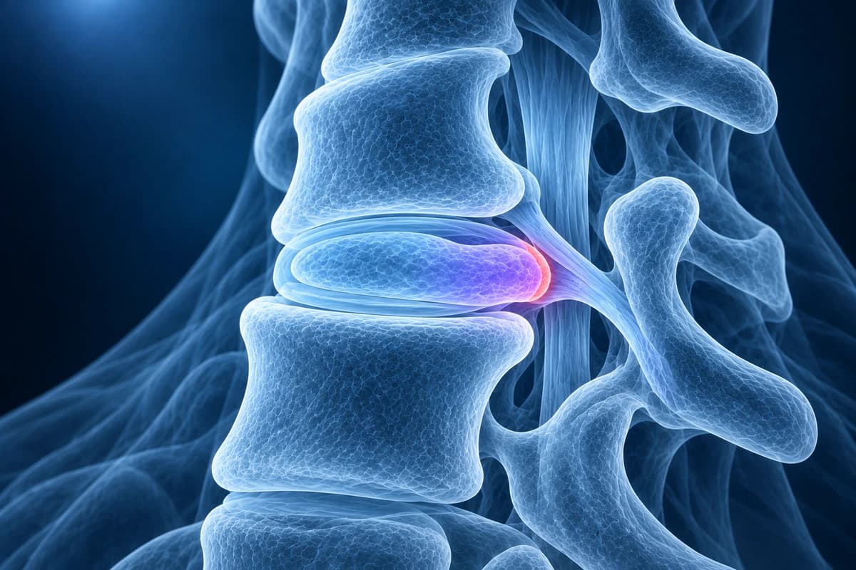
A cervical disc herniation is disc material from one of the neck discs pushing backward past its normal edge, toward a nerve root or the spinal cord. It is most often seen at C5–C6 and C6–C7. Many herniations seen on MRI cause no symptoms, and most painful ones that only affect a nerve root settle without surgery over weeks to months.
Common Symptoms
- Sharp or burning pain that travels from the neck over the shoulder blade and down one arm
- Pins and needles or numbness in a strip of the forearm, thumb, index or middle finger
- Arm pain that eases when the hand of the painful side is rested on top of the head
- Weakness lifting the arm, bending the elbow, straightening the elbow or gripping
- Neck pain and stiffness that worsen when looking up or turning the head toward the painful side
- Less commonly, clumsy hands or unsteady walking when a central disc presses on the spinal cord
When does a herniated neck disc need urgent care?
Seek same-day medical assessment if arm or hand weakness is getting worse, if both arms are affected, if your hands have become clumsy (dropping things, trouble with buttons or writing), if your walking or balance has changed, or if you notice new bladder or bowel problems. These can mean the disc is pressing on the spinal cord, not just a nerve root. Neck pain after a fall, a car crash or a sports collision, or neck pain with fever, a history of cancer or a weakened immune system, also needs prompt medical review rather than waiting for a scan explanation.
What happens when a neck disc herniates?
The seven neck vertebrae are separated by six discs, from C2–C3 down to C6–C7, plus the junction disc at C7–T1. Each disc has a tough outer ring and a softer centre. With age and repeated loading the ring develops small tears, and part of the centre can squeeze through. In the neck the space is tight: the spinal canal is narrower than in the lower back, and each nerve root leaves through a short bony tunnel called the neural foramen. A few millimetres of displaced disc can therefore matter more here than it would lower down.
Two patterns follow. A herniation that drifts to the side into the foramen irritates one nerve root and causes arm pain and tingling, known as cervical radiculopathy. A herniation in the middle of the canal can flatten the front of the spinal cord and, if large enough, cause cervical myelopathy, a slower but more serious problem with hand dexterity and walking. Younger adults more often have a soft disc herniation after a strain; people over about 50 more often have a hard ridge of disc and bone spur, part of cervical spondylosis.
Which nerve does a C5–C6 or C6–C7 herniation affect?
In the neck, a nerve root exits above the vertebra of the same number, so the disc below a vertebra usually presses on the root one number lower. A C5–C6 herniation typically affects the C6 root: pain into the thumb side of the forearm, numbness of the thumb and index finger, weaker elbow bending and wrist extension, and a reduced brachioradialis reflex. A C6–C7 herniation typically affects the C7 root: pain down the back of the arm into the middle finger, weaker elbow straightening and a reduced triceps reflex. A C4–C5 herniation tends to involve the shoulder and the deltoid, and C7–T1 the ring and little fingers and the small hand muscles.
These maps are a guide, not a rule; nerve territories overlap and vary between people. That is why a clinician compares the scan with the examination before blaming a particular level. Our explainer on C5–C6 and C6–C7 disc herniation walks through the two common levels with their typical symptoms side by side.
What does my cervical MRI report mean?
- "Right paracentral disc protrusion at C5–C6"
- Disc material extends a little past the vertebral edge, just off the midline on the right. A protrusion has a broad base; it is the most common and usually the least dramatic type of herniation.
- "Foraminal disc extrusion"
- A piece of disc has pushed further out through a narrow neck into the nerve root tunnel. Extrusions are more likely to cause arm pain, but they also tend to shrink over time as the body reabsorbs them.
- "Effaces the ventral CSF" or "contacts the ventral cord"
- The fluid layer in front of the spinal cord is pushed away, or the disc touches the cord. Touching is not the same as squeezing; "deforms" or "flattens the cord" describes true compression.
- "Disc osteophyte complex"
- A combined ridge of bulging disc and bone spur. It is a wear-related hard disc rather than a fresh soft herniation. See our disc osteophyte complex explainer.
- "Moderate left foraminal narrowing"
- The tunnel for the exiting root is reduced in size. Reports usually grade it mild, moderate or severe; the side and level should match the arm symptoms to be relevant.
- "T2 hyperintense cord signal"
- A bright spot inside the cord on T2-weighted images at the level of compression. It suggests the cord itself is stressed and is one of the findings surgeons weigh most heavily.
More wording is decoded in cervical MRI report phrases explained and in how to read a cervical spine MRI.
Do you need an MRI for a herniated disc in the neck?
Not always at first. The ACR Appropriateness Criteria for cervical neck pain or radiculopathy treat imaging as usually unnecessary early on when there are no red flags and no neurological deficit, because most episodes improve with time and conservative care. MRI without contrast becomes the test of choice when arm symptoms persist despite several weeks of treatment, when there is weakness or signs of cord involvement, or when injections or surgery are being considered. Neck X-rays show alignment, disc height and bone spurs but cannot show the disc pressing on a nerve.
CT is useful for bone: it separates a hard spur from soft disc and is used with myelography when MRI is not possible, for example with some pacemakers. Nerve conduction studies and EMG are sometimes added when it is unclear whether the problem is in the neck or in a nerve further down the arm, such as carpal tunnel syndrome. Our guide do you need an MRI for neck pain? covers the timing question in more detail, and CT vs MRI for cervical radiculopathy compares the two scans.
What we look for on a cervical disc MRI
- The level and side of the herniation, and whether it is central, paracentral or foraminal
- Protrusion versus extrusion, and any free fragment that has migrated above or below the disc
- Whether the disc only touches the cord or flattens it, and whether CSF is still visible around the cord
- Bright T2 signal within the cord at the compressed level
- Foraminal narrowing on the axial images, matched to the side of arm symptoms
- Bone spurs, uncovertebral hypertrophy and loss of disc height at neighbouring levels
Is a disc bulge on my neck MRI normal?
Often, yes. In a Japanese study of 1,211 adults with no neck or arm symptoms, disc bulging on MRI was found in most participants, and it became more common with each decade of age; some even showed the cord being indented. The lesson is that a finding on the report is only meaningful when it fits the story: the right level, the right side and the right nerve. A report listing bulges at four levels in someone with symptoms down one arm usually points to one level that matters and several that are background change.
If your report also mentions "straightening of the cervical lordosis", that is a common, usually non-specific finding linked to muscle spasm or positioning in the scanner; see what straightening of the cervical lordosis means.
How is a herniated disc in the neck treated?
Most people with arm pain from a cervical disc and no cord compression start with conservative care: staying active within comfort, pain relief, a short course of other medicines chosen by the treating doctor, and physiotherapy focused on posture, neck and shoulder-blade strength and gentle nerve mobility. A systematic review of the natural course found that many patients improve substantially over the following months. An image-guided epidural or nerve root injection is sometimes offered when pain is severe and not settling.
Surgery is considered for progressive weakness, signs of myelopathy, or disabling arm pain that has not responded to an adequate trial of non-surgical care. Common operations are anterior cervical discectomy and fusion (removing the disc from the front and fusing the level), cervical disc replacement in selected patients, and posterior foraminotomy for a soft herniation sitting in the side tunnel. If you have already had an operation, our guide to MRI after cervical fusion explains how hardware and the levels next to it are reported.
How is a neck disc herniation different from one in the lower back?
The disc behaves the same way, but the consequences differ. In the lower back the canal below the cord holds only nerve roots, so the main worries are sciatica and, rarely, cauda equina syndrome. In the neck the spinal cord itself runs through the canal, so a large central herniation can affect both arms and the legs. Neck symptoms map to the arm and hand rather than the leg, and the nerve numbering rule is different. If you are reading about a lumbar disc as well, keep the two reports separate; terms like "contacts the cord" only apply to the neck and upper back.
Can I look at my own neck MRI?
You can open your scan privately in the free MRI viewer; the files stay on your device. Scroll the sagittal images to find the level named in your report, then switch to the axial slices through that disc to see which side the disc points to. For a plain-language explanation of your images and report, upload them for an AI review. It explains findings; it is not a diagnosis and does not replace your radiologist, spine surgeon or physician.
Frequently Asked Questions
Can a herniated disc in the neck heal on its own?
Many do improve without surgery. Larger extruded fragments are often partly reabsorbed by the body, and arm pain frequently eases over weeks to months even when the MRI still shows some disc. Worsening weakness or signs of cord compression are the exceptions that need a specialist opinion promptly.
Which is worse, a C5–C6 or a C6–C7 herniation?
Neither level is worse in itself. What matters is the size and direction of the herniation, whether it compresses the cord or a single root, and how much strength or sensation is lost. C5–C6 tends to affect the thumb side and elbow bending; C6–C7 the middle finger and elbow straightening.
What does "contacts the spinal cord" mean on a neck MRI?
It means the disc reaches the surface of the cord without clearly changing its shape. It is less serious than "compresses" or "flattens" the cord, and much less concerning than a bright cord signal. It should still be read alongside your examination, especially if you have any hand clumsiness or balance change.
Why does lifting my arm over my head ease the pain?
Resting the hand on the head slackens the irritated nerve root, so many people with a cervical disc herniation find relief that way. Clinicians call it the shoulder abduction relief sign, and it points toward a nerve root problem in the neck rather than a shoulder problem.
Can a neck disc herniation cause headaches?
Lower neck herniations mainly cause neck, shoulder-blade and arm pain. Headaches arising from the neck usually come from the upper joints and nerves, from C1 to C3; our page on cervicogenic headache explains how that is diagnosed.
Sources
- Expert Panel on Neurological Imaging; McDonald MA, Kirsch CFE, et al. ACR Appropriateness Criteria Cervical Neck Pain or Cervical Radiculopathy. J Am Coll Radiol. 2019;16(5S):S57-S76.
- Bono CM, Ghiselli G, Gilbert TJ, et al. An evidence-based clinical guideline for the diagnosis and treatment of cervical radiculopathy from degenerative disorders. Spine J. 2011;11(1):64-72.
- Nakashima H, Yukawa Y, Suda K, et al. Abnormal findings on magnetic resonance images of the cervical spines in 1211 asymptomatic subjects. Spine (Phila Pa 1976). 2015;40(6):392-398.
- Wong JJ, Côté P, Quesnele JJ, Stern PJ, Mior SA. The course and prognostic factors of symptomatic cervical disc herniation with radiculopathy: a systematic review of the literature. Spine J. 2014;14(8):1781-1789.
Related Articles
Why C5-C6 and C6-C7 are the neck discs that herniate most, which nerve root each one pinches, how the arm symptoms differ, and what protrusion, extrusion and foraminal mean on your MRI.
What an MRI can and cannot show after an anterior cervical fusion (ACDF): metal artifact, adjacent segment disease, residual compression and cord signal, when CT or X-ray answers better, and which symptoms need urgent care.
How a herniated disc in the neck recovers phase by phase, how often herniations shrink on their own, when a repeat MRI helps, and which signs change the plan.
Understand your imaging before your next appointment
MRI, X-ray or CT from a CD, folder or ZIP. A PDF report or photos of the films work too.
Free to open and preview, no account needed. An optional AI report from $10 explains your images in plain language, with questions for your doctor.
Your raw files stay on this device. Only rendered images and minimal context are sent when you request an AI report.
Medical Disclaimer: This page is for informational and educational purposes only. It does not constitute medical advice, diagnosis, or treatment. AI-generated analysis may contain errors. Always consult a qualified healthcare professional for medical decisions. Full Disclaimer