
Discitis-Osteomyelitis: Spine Infection on MRI and When to Get Urgent Care
Discitis-osteomyelitis, also called spondylodiscitis or vertebral osteomyelitis, is an infection of an intervertebral disc and the two vertebrae on either side of it. It is uncommon but serious. MRI of the spine with and without contrast is the key imaging test, blood cultures or a biopsy identify the germ, and treatment is usually weeks of antibiotics, with surgery for abscess, instability or nerve compression.
Some signs described on this page need prompt medical care. If you have them, get care first. Reading about imaging can wait.
Common Symptoms
- Constant neck or back pain that keeps getting worse over days to weeks and does not settle with rest
- Pain that is bad at night and wakes you from sleep
- Fever, chills or night sweats, although many people with spine infection have no fever
- Marked tenderness when the affected part of the spine is pressed or tapped
- New weakness, numbness or tingling in the arms or legs, or difficulty walking
- Painful or difficult swallowing when the infection is in the neck
When should you go to the emergency department?
Go to an emergency department today, or call emergency services, if you have worsening neck or back pain together with any of these: fever or chills; current or past injecting drug use; a spinal operation, injection, epidural or nerve block in recent weeks or months; a weakened immune system from diabetes, dialysis, cancer treatment, steroids, transplant medicines or HIV; a recent bloodstream, urinary or skin infection, or a heart valve infection; or any new weakness, numbness, unsteady walking, or bladder or bowel change. Spine infection can spread into the space around the spinal cord and cause permanent paralysis within hours to days. Do not wait for a routine appointment or an online report review.
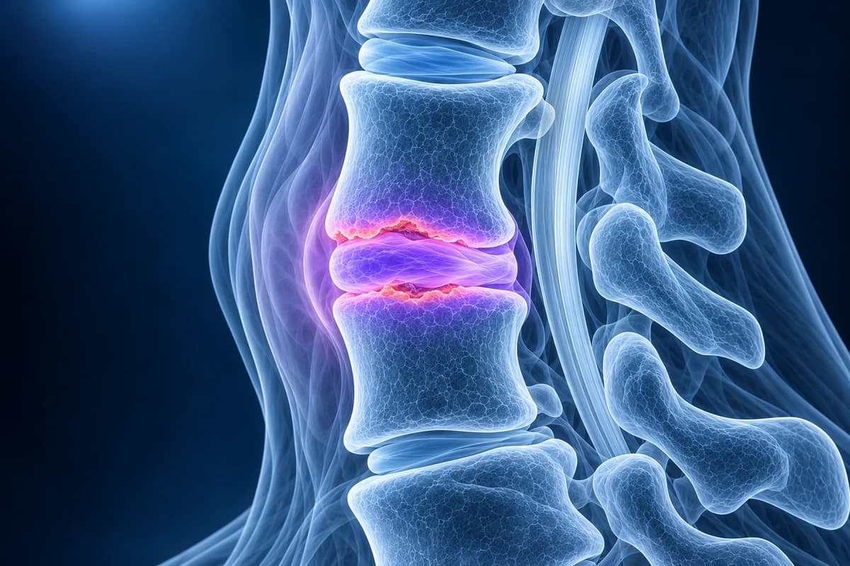
How does a spine infection start?
In adults, bacteria most often reach the spine through the bloodstream from an infection elsewhere, such as the skin, urinary tract, a heart valve, an intravenous line or injecting equipment. The bone just under the endplates has a rich blood supply, so germs lodge there first and then spread into the disc, which has almost no blood supply of its own and cannot easily fight infection. Less often, bacteria are introduced directly during spine surgery or an injection, or spread from a nearby infection such as a throat abscess.
Staphylococcus aureus is the most frequent cause. Tuberculosis and brucellosis are important causes in some regions and in people who have lived in or travelled there, and fungi are seen mainly in people with very weak immune defences. The lumbar spine is affected most often, then the thoracic spine. The neck is the least common site, but a cervical infection is the most dangerous because the canal is narrow, the spinal cord is close, and pus can track into the tissues behind the throat.
Why is spine infection so often diagnosed late?
The first symptom is usually just pain, which is easy to mistake for a strain or ordinary wear. Fever is often absent, especially in older adults or people already taking antibiotics or painkillers that lower temperature, and early X-rays look normal. In a study of emergency department patients with spinal epidural abscess, the textbook combination of back pain, fever and a neurological deficit was present in only a minority at first visit, and most patients experienced a delay in diagnosis, with worse outcomes when weakness had already developed.
The practical message is simple: steadily worsening, constant spine pain in someone with a risk factor needs blood tests and urgent MRI, even without fever. Blood markers of inflammation, the ESR and CRP, are raised in most cases and are also used to track the response to treatment.
Which scan shows a spine infection?
MRI of the affected region with and without gadolinium contrast is the test of choice in both the IDSA guideline on native vertebral osteomyelitis and the ACR Appropriateness Criteria for suspected spine infection. It shows the infected disc and bone early, and contrast outlines abscesses in the epidural space and the muscles beside the spine. Because infection can involve more than one level, the radiologist may extend the scan to other parts of the spine.
Plain X-rays are often normal for the first two to three weeks before disc space narrowing and blurred endplates appear, so a normal X-ray does not exclude infection. CT shows bone destruction well and is used to guide a needle biopsy. When MRI is not possible, nuclear medicine scans such as FDG-PET/CT are alternatives. Our guide on how to read a cervical spine MRI explains the T1, T2 and STIR sequences mentioned in these reports.
What does a discitis-osteomyelitis MRI report mean?
- "T2 hyperintense disc with fluid signal"
- The disc looks unusually bright on water-sensitive images, as if filled with fluid. A worn disc normally turns dark, so a bright, swollen disc raises concern for infection.
- "Endplate erosion" or "endplate destruction"
- The thin bone surfaces above and below the disc are irregular or partly eaten away. Damage on both sides of one disc is the classic pattern of discitis-osteomyelitis.
- "Marrow edema with T1 hypointensity of the adjacent vertebral bodies"
- The bone marrow next to the disc has lost its normal fatty signal and holds extra fluid, which is how infected bone looks on MRI.
- "Paravertebral or prevertebral phlegmon"
- Inflamed, swollen soft tissue beside or in front of the spine. A phlegmon enhances solidly with contrast; an abscess has a liquid centre with an enhancing rim.
- "Rim-enhancing epidural collection"
- An epidural abscess inside the spinal canal. If it presses on the cord or nerve roots, it is a surgical emergency.
- "Findings may represent Modic type 1 change; correlate for infection"
- Degenerative endplate inflammation can look similar to early infection. The report is flagging uncertainty, which is resolved with blood tests, contrast imaging and sometimes biopsy, not by waiting.
For more report wording, see cervical MRI report phrases explained.
What we look for on suspected spine infection imaging
- Bright, fluid-like signal in the disc with loss of disc height
- Erosion of both endplates around the same disc, with marrow edema in the two vertebrae
- Enhancement of the disc and bone after contrast
- Epidural, paravertebral or prevertebral collections and their effect on the spinal cord or airway
- Vertebral collapse, kyphosis or instability at the infected level
- Additional infected levels elsewhere in the spine
What else can look like a spine infection on MRI?
Several conditions mimic infection. Active degenerative endplate change, called Modic type 1, causes marrow edema on both sides of a worn disc, but the disc itself is usually dark and the endplates intact. Gas in the disc, the "vacuum phenomenon", generally points toward degeneration rather than infection. Tumours tend to involve the vertebral body and spare the disc. Fractures, inflammatory arthritis, and spine changes in long-term dialysis or in nerve-damaged joints can also confuse the picture. In the neck, cervical spondylosis with an active endplate reaction is a common source of "cannot exclude infection" wording. When doubt remains, a CT-guided biopsy settles it.
Tuberculous spondylitis has its own pattern: it often spreads under the ligaments to several vertebrae, may relatively spare the disc until late, and can form large, thin-walled abscesses with little pain. It needs a different and longer treatment, which is one reason confirming the organism matters.
How is discitis-osteomyelitis treated?
Treatment depends on identifying the germ. Doctors take blood cultures first; if they are negative, an image-guided needle biopsy of the disc or bone usually follows. The IDSA guideline suggests holding antibiotics until a sample is taken in people who are stable and have no neurological deficit, because starting early can make the germ impossible to grow. People with sepsis, low blood pressure or new weakness are treated immediately. For most bacterial infections the guideline recommends a total of about six weeks of antibiotics, often starting intravenously; tuberculosis, fungal infection and infections around implants need longer courses.
Surgery is needed for an epidural abscess pressing on the cord, progressive neurological deficit, spinal instability or collapse, or infection that does not respond to antibiotics. In the neck this usually means an anterior operation to clean out the infection and rebuild the column, sometimes with posterior fixation. Recovery is tracked with symptoms, examination and falling inflammatory markers. MRI often looks worse for weeks even when treatment is working, so repeat scans are mainly used when a patient is not improving or develops new symptoms.
Can infection develop after neck surgery?
Yes. Infection after spine surgery is uncommon but most often shows up as wound redness or drainage, fever, or neck pain that gets worse instead of better in the weeks after an operation. Any of these after a fusion or disc replacement should be reported to the surgical team the same day. Metal implants create artefact on MRI, which is covered in cervical MRI after fusion. Other urgent neck pages include cervical myelopathy and cervical spine fracture.
Can I review my spine MRI myself?
If you are already under care and want to understand an existing scan, you can open it privately in the free MRI viewer; files stay on your device. You can also upload images and the report for a plain-language AI explanation. It explains wording and findings; it is not a diagnosis, cannot rule out infection, and must never delay emergency care if you have any of the warning signs above.
Frequently Asked Questions
Can you have a spine infection without a fever?
Yes. Many people with vertebral osteomyelitis have no fever when they first seek help. Constant, worsening spine pain with a risk factor such as injecting drug use, a recent spinal procedure, diabetes, dialysis or a weakened immune system should prompt urgent assessment even with a normal temperature.
Why does the MRI need contrast?
Contrast shows which tissue is actively inflamed and separates a solid phlegmon from a liquid abscess that may need draining. It is especially important for finding epidural abscesses that can compress the spinal cord.
Is discitis the same as osteomyelitis?
Discitis is infection of the disc; vertebral osteomyelitis is infection of the bone. In adults they almost always occur together, so the combined term discitis-osteomyelitis, or spondylodiscitis, is used.
How long does recovery take?
Antibiotic treatment commonly lasts around six weeks or longer, and pain and stiffness can take months to settle. The infected level often fuses on its own as it heals. Outcomes are best when the infection is found before nerve damage develops.
My report says "cannot exclude discitis". What should I do?
Contact the doctor who ordered the scan the same day. That wording means the radiologist sees features that could be infection and wants it ruled in or out, usually with blood tests, contrast MRI if not already done, and sometimes a biopsy. If you have fever, weakness or worsening pain, go to an emergency department.
Sources
- Berbari EF, Kanj SS, Kowalski TJ, et al. 2015 Infectious Diseases Society of America (IDSA) clinical practice guidelines for the diagnosis and treatment of native vertebral osteomyelitis in adults. Clin Infect Dis. 2015;61(6):e26-e46.
- Zimmerli W. Clinical practice. Vertebral osteomyelitis. N Engl J Med. 2010;362(11):1022-1029.
- Davis DP, Wold RM, Patel RJ, et al. The clinical presentation and impact of diagnostic delays on emergency department patients with spinal epidural abscess. J Emerg Med. 2004;26(3):285-291.
- Expert Panel on Neurological Imaging; Ortiz AO, Levitt A, et al. ACR Appropriateness Criteria Suspected Spine Infection. J Am Coll Radiol. 2021;18(11S):S488-S501.
Related Articles
When a cervical MRI needs gadolinium (infection, tumour, scar after surgery, MS) and when it does not, plus kidney, retention, allergy and pregnancy safety.
What sets the price of a cervical spine MRI: hospital or imaging centre, contrast (CPT 72141 vs 72156), 1.5T or 3T, insurance, prior authorisation and cash pay.
Most neck pain does not need an MRI. Learn the warning signs that do call for imaging, why X-ray or CT comes first after an injury, and how to read the age-related findings a neck MRI almost always shows.
Do not delay urgent medical care to use this tool.
After you get care, understand your imaging
MRI, X-ray or CT from a CD, folder or ZIP. A PDF report or photos of the films work too.
Free to open and preview, no account needed. An optional AI report from $10 explains your images in plain language, with questions for your doctor.
Your raw files stay on this device. Only rendered images and minimal context are sent when you request an AI report.
Medical Disclaimer: This page is for informational and educational purposes only. It does not constitute medical advice, diagnosis, or treatment. AI-generated analysis may contain errors. Always consult a qualified healthcare professional for medical decisions. Full Disclaimer