
Do You Need an MRI for Neck Pain? When Imaging Helps and When It Does Not
Most neck pain does not need an MRI. Learn the warning signs that do call for imaging, why X-ray or CT comes first after an injury, and how to read the age-related findings a neck MRI almost always shows.
Key Takeaways
- Most neck pain without neurological symptoms or red flags does not need an MRI at first
- Progressive weakness, clumsy hands, unsteady walking, bladder or bowel change, fever or major trauma need prompt medical care before any booked scan
- After an injury, NEXUS or the Canadian C-spine rule decides whether to image, and CT usually comes first
- Disc bulging appeared in 87.6% of symptom-free adults in a large MRI study, so findings must be matched to symptoms
- MRI is most useful for persistent nerve symptoms, suspected cord compression and planning treatment
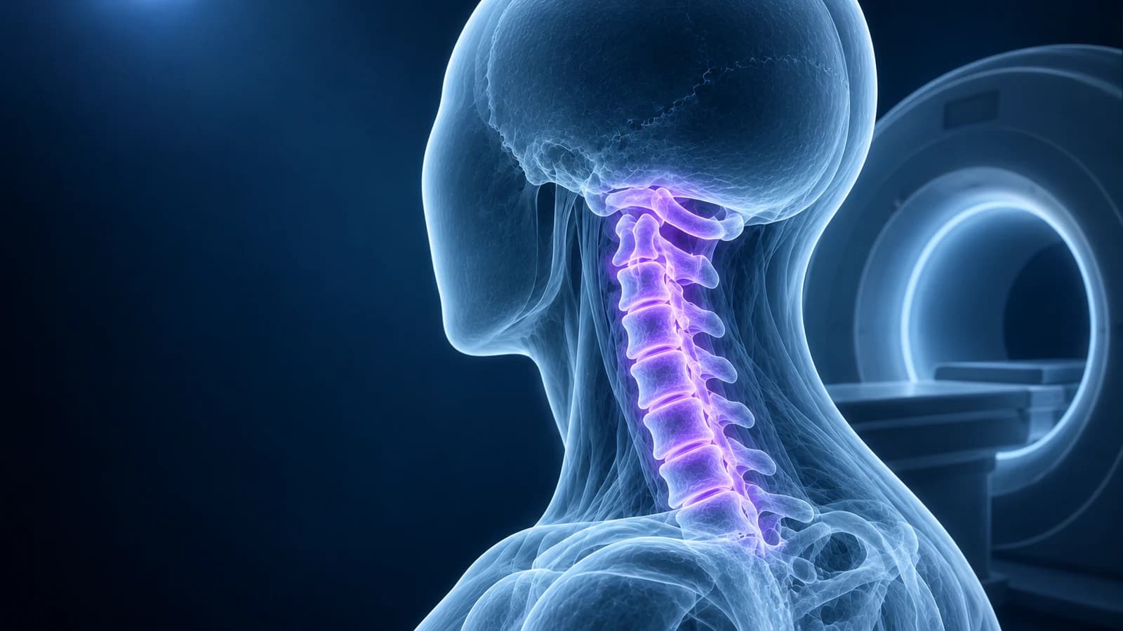
Most people with neck pain do not need an MRI. Neck pain without arm weakness, numbness, clumsy hands, balance problems, fever or a significant injury usually improves over weeks with movement and simple treatment, and an early scan rarely changes that plan. MRI becomes the right test when nerve or spinal cord symptoms appear, when pain persists despite about six weeks of care, or when a warning sign is present.
The reason doctors hold back is not cost alone. A cervical MRI in an adult almost always finds something: bulging discs, small spurs, thinning discs, a little narrowing of a nerve opening. Those findings are common in people with no neck pain at all, so a scan taken too early can attach a frightening label to an ordinary, self-limiting episode. This guide explains when imaging helps, which test comes first in which situation, and how to read the report if you already have one. It is general education, not a diagnosis.
Which neck symptoms need urgent care rather than a booked scan?
Seek same-day medical care, and an emergency department if symptoms are severe or sudden, if neck pain comes with any of the following. These are reasons to be examined first; the clinician decides which imaging, if any, follows.
- Weakness in an arm or hand that is getting worse, or numbness spreading across both arms
- Clumsy hands, dropping things, trouble with buttons or writing, or a new unsteady or wide-based walk
- New difficulty controlling or starting urination, or loss of bowel control
- Neck pain after a fall, a road collision, a diving accident or any significant blow to the head
- Fever, chills or feeling unwell with neck pain, especially after an infection or injecting drugs
- A history of cancer, weakened immunity, unexplained weight loss or pain that is worse at night and at rest
- A sudden severe headache with a stiff neck, or neck stiffness with confusion, a rash or sensitivity to light
Why is an MRI not the first step for ordinary neck pain?
The American College of Radiology Appropriateness Criteria for cervical neck pain separate patients by whether there are red flags, whether there is radiculopathy, and how long symptoms have lasted. For neck pain without red flags, imaging is generally not needed at the start, and when a first picture is wanted for chronic pain, plain X-rays are the usual starting point. MRI is reserved for the situations where it can actually answer a question.
The other half of the reasoning is what a scan finds in people who feel fine. In a study of 1,211 adults with no neck symptoms, 87.6% had disc bulging on MRI, including most volunteers in their twenties. Compression touching the spinal cord was found in 5.3% and a bright cord signal in 2.3%, both becoming more common after age 50. Those numbers are the background noise against which your own scan is read, and they explain why a report full of findings can still belong to a neck that will recover.
When does a neck MRI actually help?
- Arm pain, tingling or weakness in a nerve-root pattern that persists or worsens after several weeks of care, or that is severe from the outset
- Any sign of spinal cord involvement, such as clumsy hands, balance change or brisk reflexes found on examination
- Red flags for infection, tumour or inflammatory disease, where MRI, often with contrast, shows marrow and soft tissue that X-ray cannot
- Neurological symptoms after an injury, or a CT that leaves a ligament or cord question open
- Planning a targeted injection or surgery, when the exact level and cause of compression matter
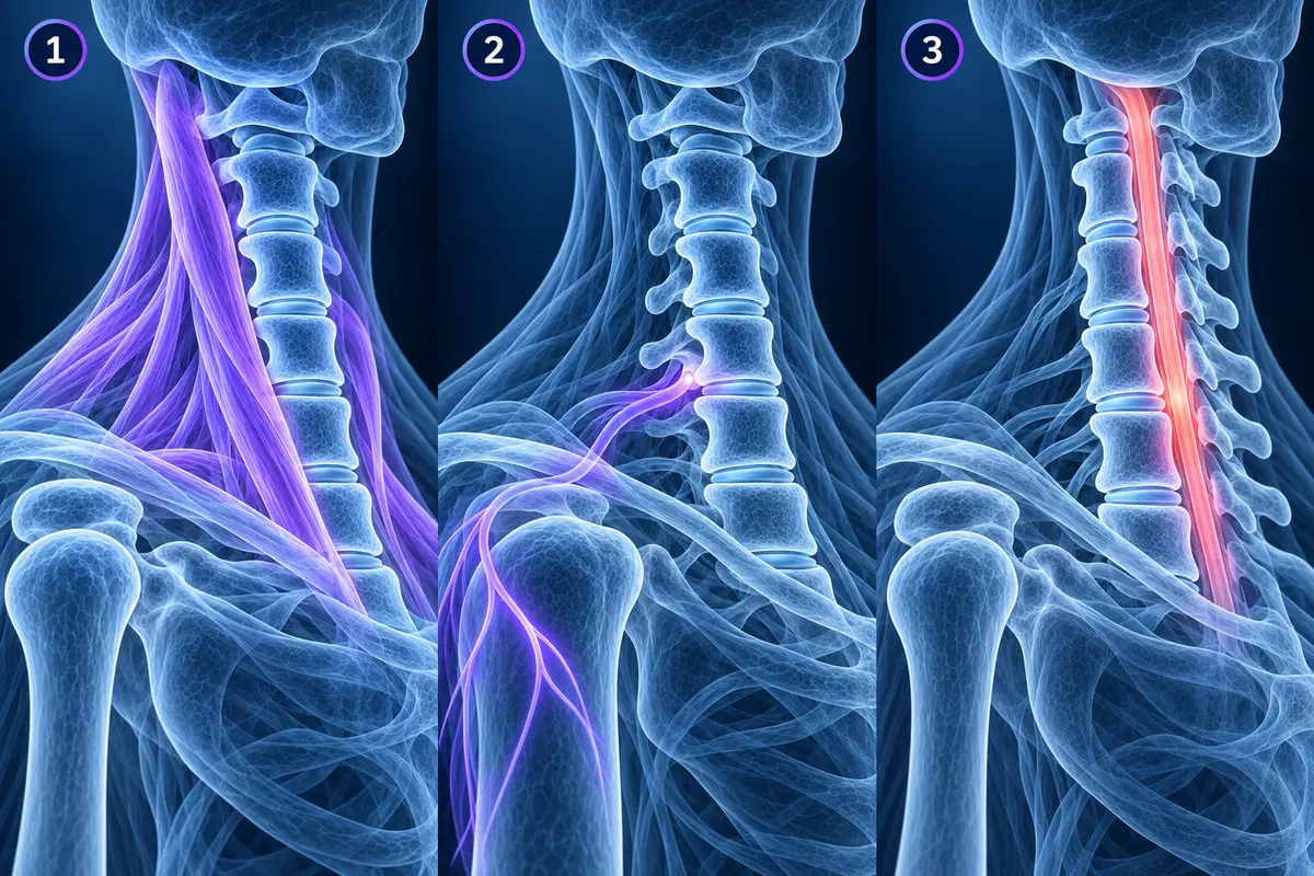
Pain that radiates down one arm along a band, often with tingling in particular fingers, suggests a pinched nerve root, described in our guide to cervical radiculopathy. Many of these settle without a scan. Hand clumsiness and a change in walking suggest pressure on the cord itself, which is a different and more time-sensitive problem covered in cervical myelopathy warning signs on MRI.
Do you need imaging after a neck injury?
After a car crash or a fall, the first question is whether the bones could be broken, and emergency clinicians answer it with validated decision rules rather than routine scanning. The NEXUS criteria clear the neck without imaging when all five are true: no tenderness over the midline of the back of the neck, no intoxication, normal alertness, no focal neurological deficit and no painful distracting injury elsewhere.
The Canadian C-spine rule, designed for alert and stable adults, sends anyone aged 65 or older, anyone with a dangerous mechanism such as a high-speed or rollover collision or a fall from height, and anyone with tingling in the limbs straight to imaging. Lower-risk patients, for example after a simple rear-end collision, who can sit up and walk and have no midline tenderness, are then asked to turn the head 45 degrees to each side; if they can, imaging is not needed.
When an adult does meet the criteria, the ACR guidance for suspected spine trauma favours CT over plain X-rays as the first test, because CT is faster and far better at finding fractures. MRI is added for neurological symptoms or when a ligament injury is suspected. Our pages on whiplash injury and what a whiplash MRI shows cover the soft-tissue side of the story.
Is an X-ray, CT or MRI the right first test?
Each test answers a different question. A cervical spine X-ray shows alignment, disc height and bone spurs in a few seconds, and flexion and extension views add movement, which is the only way to see a slip that appears when the head bends. CT maps bone in fine detail, which suits fractures and the bony narrowing of nerve openings. MRI is the only one of the three that shows the discs, nerve roots, spinal cord, ligaments and bone marrow directly.
For a pinched nerve, the choice between the two cross-sectional tests is not always obvious, and our comparison of CT vs MRI for cervical radiculopathy explains when each is preferred. People who cannot have an MRI, for example because of some implanted devices, can often have a CT or a CT myelogram instead.
What will a neck MRI probably show if you are over 40?
Expect the report to describe wear somewhere between C3 and C7, most often at C5–C6 and C6–C7, the levels that bend the most. The wording is standardised, and most of it describes ageing rather than injury.
- “Mild multilevel degenerative change” or “spondylosis” — general wear of the discs and small joints, usual for age and described in cervical spondylosis
- “Disc osteophyte complex” — a bulging disc edge and bone spur merged into one ridge; the report should say whether it touches the cord or a nerve
- “Uncovertebral hypertrophy with foraminal narrowing” — spurs from the small side joints crowding the opening where a nerve root exits
- “Straightening of the cervical lordosis” — the normal forward curve looks flatter, frequently a matter of posture or muscle spasm on the day of the scan
- “Effacement of the ventral CSF” — the fluid in front of the cord is pressed thin, which is less than cord compression
- “T2 hyperintense cord signal” — a bright spot inside the spinal cord itself, the finding that deserves the closest follow-up
For the full glossary, see cervical MRI report phrases explained and our walk-through of how to read a cervical spine MRI. If you would like to scroll through your own sagittal and axial slices, the free MRI viewer opens DICOM files in your browser without uploading them.
What usually happens if you wait instead of scanning?
Waiting does not mean doing nothing. A typical first plan combines staying active within comfort, short courses of pain relief that suit you, and guided exercise or physiotherapy for posture, mobility and shoulder-blade strength. A pinched nerve in the neck often settles over weeks to a few months as inflammation around the root calms and a soft disc fragment shrinks, which is why reviews of cervical radiculopathy put non-operative care first for most patients.
The waiting period has conditions. If weakness develops, symptoms spread to both arms or the legs, or the pain is unbearable despite treatment, the plan changes and imaging moves forward. A scan is also reasonable once a specialist is considering an injection or an operation, because those decisions depend on the exact level and type of compression.
Frequently Asked Questions
How long should I wait before asking for an MRI for neck pain?
Without warning signs, guidelines commonly suggest around six weeks of conservative care before imaging is reconsidered, because most episodes improve in that time. That timeline is replaced immediately by prompt assessment if weakness, clumsy hands, walking problems, bladder or bowel change, fever or a significant injury are present.
Can an X-ray show a pinched nerve in the neck?
Not directly. An X-ray shows the bones, disc height and spurs, and an oblique view can show a narrowed nerve opening, but nerves and discs themselves are invisible on X-ray. MRI shows the nerve root and what is pressing on it, which is why it is the usual test when a pinched nerve is not improving.
Does a neck MRI need contrast?
Usually not. A standard cervical MRI for neck pain or a pinched nerve is done without contrast. Gadolinium contrast is added when infection, a tumour, an inflammatory condition of the cord or a post-operative question is being investigated.
My MRI shows disc bulges at several levels. Is that why my neck hurts?
Possibly, but not necessarily. Multilevel disc bulging is so common in adults without symptoms that it cannot be blamed on its own. A clinician links a finding to your pain when its level matches the arm pattern, the examination and the timeline. Findings that touch a nerve root on the painful side or press on the cord carry more weight than a bulge in empty space.
Is a 3T MRI better for the neck than a 1.5T MRI?
A 3T scanner gives sharper detail of nerve roots and small structures, but a well-performed 1.5T cervical MRI answers the usual questions about discs, nerve openings and the cord. The quality of the protocol and the reader matters more than the field strength for most neck pain.
Sources
- Expert Panel on Neurological Imaging; McDonald MA, Kirsch CFE, et al. ACR Appropriateness Criteria Cervical Neck Pain or Cervical Radiculopathy. J Am Coll Radiol. 2019;16(5S):S57-S76.
- Nakashima H, Yukawa Y, Suda K, et al. Abnormal findings on magnetic resonance images of the cervical spines in 1211 asymptomatic subjects. Spine (Phila Pa 1976). 2015;40(6):392-398.
- Stiell IG, Wells GA, Vandemheen KL, et al. The Canadian C-spine rule for radiography in alert and stable trauma patients. JAMA. 2001;286(15):1841-1848.
- Hoffman JR, Mower WR, Wolfson AB, et al. Validity of a set of clinical criteria to rule out injury to the cervical spine in patients with blunt trauma. N Engl J Med. 2000;343(2):94-99.
- Expert Panel on Neurological Imaging and Musculoskeletal Imaging; Beckmann NM, West OC, et al. ACR Appropriateness Criteria Suspected Spine Trauma. J Am Coll Radiol. 2019;16(5S):S264-S285.
Related Articles
What a neck X-ray can and cannot show: the standard views, the four alignment lines, disc space narrowing, bone spurs and the Torg-Pavlov ratio, and when CT or MRI is needed instead.
A plain-language guide to your neck MRI: how to count from C2 down to C7, which slices show the cord and the nerve exits, and which artifacts make a cervical scan look worse than it is.
What cord compression, effaced CSF, T2 hyperintense cord signal and myelomalacia mean on a neck MRI, which symptoms make them urgent, and how doctors weigh MRI findings against clumsy hands and balance problems.
Related Conditions
Understand your imaging before your next appointment
MRI, X-ray or CT from a CD, folder or ZIP. A PDF report or photos of the films work too.
Free to open and preview, no account needed. An optional AI report from $10 explains your images in plain language, with questions for your doctor.
Your raw files stay on this device. Only rendered images and minimal context are sent when you request an AI report.
Medical Disclaimer: This page is for informational and educational purposes only. It does not constitute medical advice, diagnosis, or treatment. AI-generated analysis may contain errors. Always consult a qualified healthcare professional for medical decisions. Full Disclaimer