
Neck MRI With vs Without Contrast: When Gadolinium Is Needed
When a cervical MRI needs gadolinium (infection, tumour, scar after surgery, MS) and when it does not, plus kidney, retention, allergy and pregnancy safety.
Key Takeaways
- Most neck MRIs for disc, nerve and stenosis questions are done without contrast
- Contrast is added for infection, tumour, scar versus disc after surgery and inflammation of the cord
- Severe allergy-like reactions to gadolinium occur in about 1 per 20,000 injections
- Kidney disease, a previous reaction and pregnancy should be mentioned before the scan
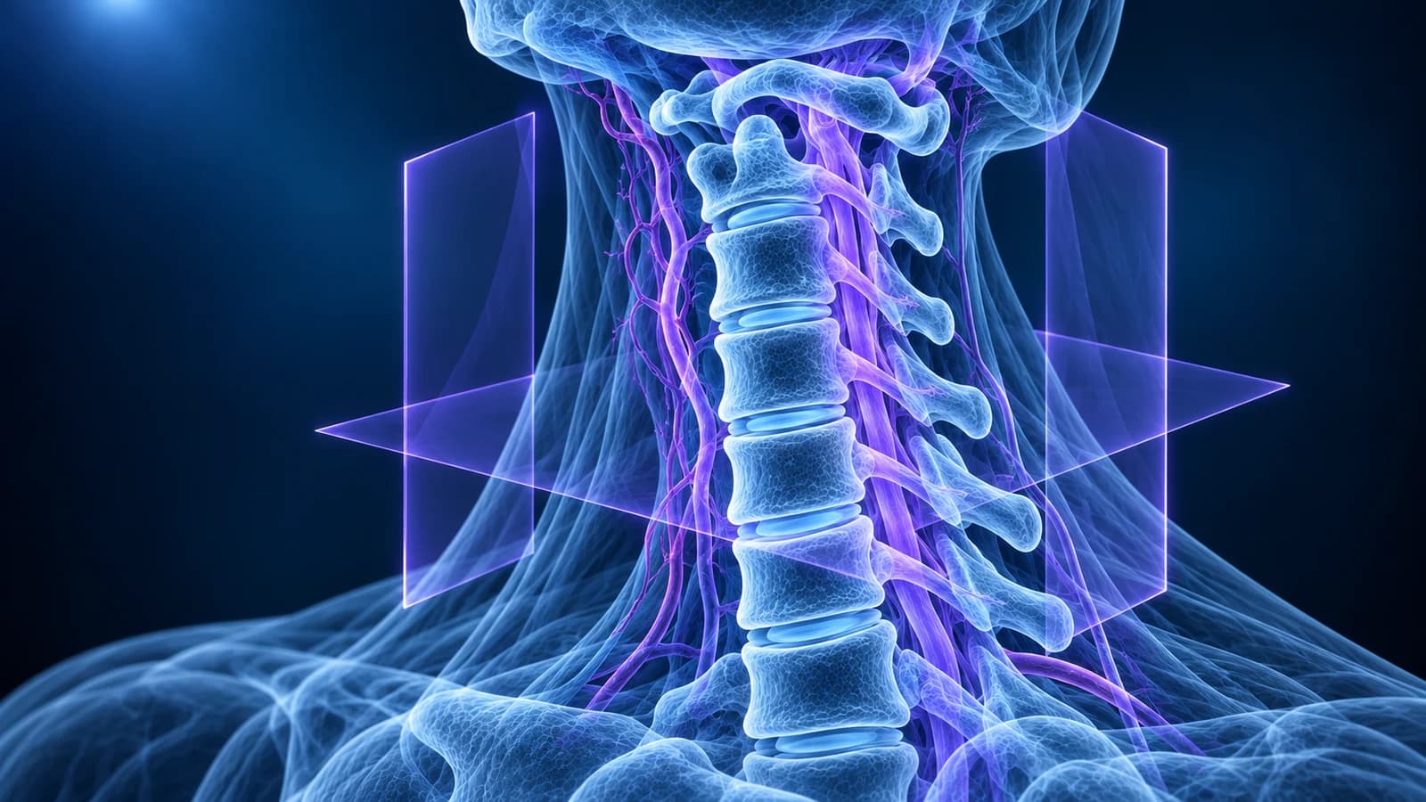
Most neck MRIs are done without contrast. A plain scan answers the usual questions about discs, bone spurs, nerve roots and cord compression. Gadolinium is added when the question is infection, a tumour, scar versus a new disc after surgery, or inflammation of the cord such as multiple sclerosis. Severe allergy-like reactions are rare, about 1 in 20,000 injections.
If your referral says “MRI cervical spine with and without contrast”, the scanner takes one set of images, a nurse or technologist injects the contrast through a small cannula in your arm, and some of the sequences are repeated. This guide explains why a doctor would ask for that, why most people do not need it, and what is known about safety. It is general education and does not replace advice from the team that ordered your scan.
What does contrast show on a cervical MRI that a plain scan cannot?
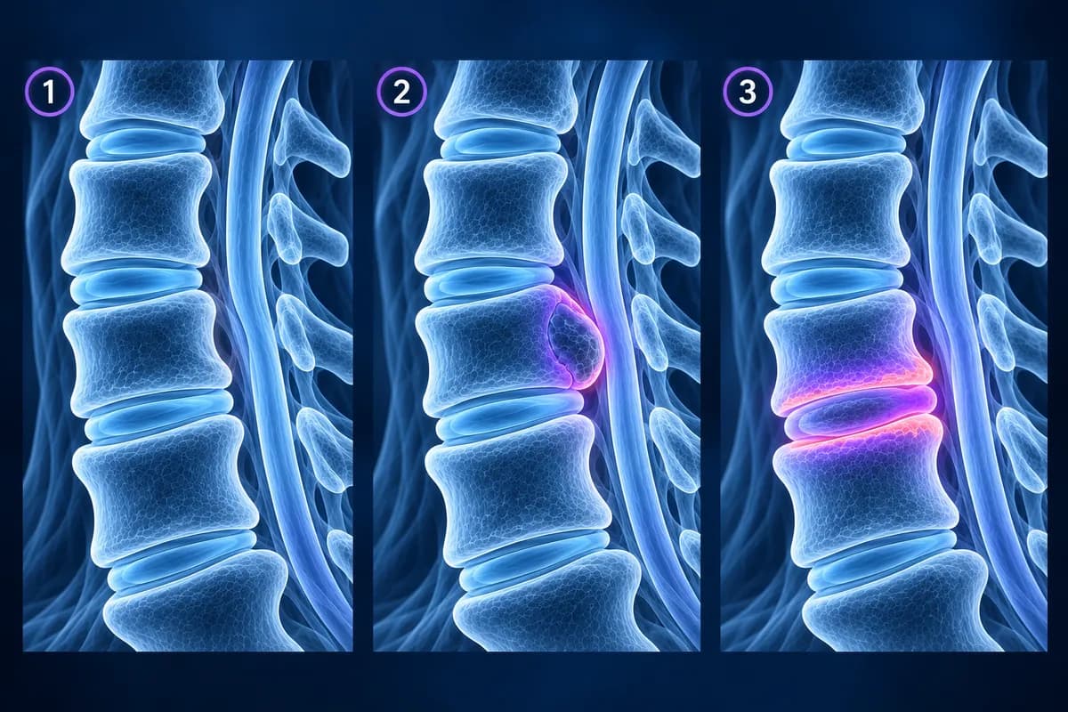
Contrast does not make the whole picture sharper. It highlights tissue by its blood supply. Healthy disc material has almost no blood vessels, so it stays dark after the injection, while inflamed or newly formed tissue takes up gadolinium and turns bright on T1-weighted images. Radiologists call this “enhancement”, and its pattern (a thin rim, a solid patch, a ring around fluid) is what separates one diagnosis from another.
That is also why contrast adds nothing to most degenerative questions. A disc bulge, a disc osteophyte complex or a narrowed nerve exit is already visible against the bright spinal fluid on T2 images, and the size of the canal does not change after an injection.
When is gadolinium added to a neck MRI?
The American College of Radiology (ACR) rates “MRI cervical spine without and with IV contrast” as usually appropriate in two neck pain situations: suspected or known infection, and known or suspected cancer. In practice four questions account for most contrast-enhanced neck scans.
- Infection: fever, raised blood markers, recent surgery, injecting drug use or a weakened immune system with new neck pain. Contrast shows how far infection has spread through the disc and bone in discitis-osteomyelitis and separates a drainable pus collection (an abscess, which enhances only at its rim) from swollen inflamed tissue.
- Tumour: a history of cancer, unexplained weight loss or night pain. Contrast helps show spread to the vertebrae or the spinal canal and outlines tumours of the cord or the nerve sheaths.
- After surgery: new arm pain at a level that was operated on. Scar tissue enhances, while a recurrent or leftover disc fragment does not, so the pre- and post-contrast images show which one is pressing on the nerve.
- Inflammation of the cord: suspected multiple sclerosis or another cause of myelitis. Lesions that are actively inflamed enhance, while older ones do not, which helps date them.
Does an MRI after neck surgery need contrast?
Not always. For new pain or arm symptoms after an operation, the ACR rates plain MRI (alongside X-rays and CT) as usually appropriate, and the contrast version as one that may be appropriate. Contrast earns its place when the team needs to know whether tissue pressing on a nerve is scar or disc. In a classic study of patients who had a second operation on the lower back, MRI before and after contrast told scar from disc correctly in 96% of the operated levels, when the scan was done six or more weeks after surgery. The same principle is applied in the neck.
Metal plates and cages cause local distortion on MRI, and the questions after a fusion, such as wear at the disc above or below, are often answered without contrast. What a post-fusion scan can and cannot show is covered in cervical MRI after fusion.
Is contrast needed for multiple sclerosis in the cervical cord?
The cervical cord is one of the places multiple sclerosis plaques are looked for, and the 2021 international MRI recommendations for MS stress the value of spinal cord imaging. At diagnosis, contrast shows whether some lesions are active at the same time as others are older, which can support the diagnosis. For routine follow-up, the same recommendations advise using gadolinium judiciously for specific questions rather than at every scan, because new lesions can usually be found by comparing plain images with the previous ones.
A bright patch inside the cord can also come from compression by a disc or a bony ridge. That pattern belongs to cervical myelopathy, sits right at the narrowed level, and is usually assessed without contrast.
When is a neck MRI without contrast enough?
For the common reasons people have a neck MRI, a plain scan is the standard. The ACR lists MRI without contrast as the usual test for long-standing neck pain with arm symptoms, the typical picture of cervical radiculopathy, and as an option for long-standing neck pain without arm symptoms. Disc herniations, bulges, foraminal narrowing, canal stenosis and wear of the small joints are all judged on the plain sequences. Whether an MRI is needed in the first place is covered in do you need an MRI for neck pain.
If a plain scan shows something unexpected, such as an enhancing-looking mass or marrow change, the radiologist may recommend a second visit with contrast. That is a targeted follow-up, not a sign that the first scan was wrong.
Is gadolinium contrast safe?
For most people, yes. More than 450 million doses had been given worldwide by 2018 with a very favourable safety record. The points worth knowing, in plain words:
- Kidneys: gadolinium leaves the body mainly through the kidneys. A rare skin and organ scarring condition (nephrogenic systemic fibrosis) was linked to older agents in people with severe kidney disease. With the agents now preferred, a 2021 ACR and National Kidney Foundation statement found zero cases after 4,931 injections in people with severely reduced kidney function, and advises weighing that very small risk against the harm of delaying a needed scan. You may be asked about kidney disease or have a blood test first.
- Retention: tiny amounts of gadolinium can remain in the brain, bone and skin for months to years. In 2017 the US FDA added a class warning about this to every gadolinium agent. No harm from retention has been proven in people with normal kidney function, and the more stable macrocyclic agents leave less behind than linear ones.
- Allergy-like reactions: a review of 716,978 injections found immediate allergy-like reactions in about 9 per 10,000, most of them not severe, and severe reactions in about 1 per 20,000. Tell the team if you reacted to gadolinium before, and mention a past reaction to iodine CT contrast too, even though it is a different drug.
- Pregnancy and breastfeeding: the ACR advises giving gadolinium during pregnancy only when the information is essential and cannot be obtained another way, so tell the team if you are or may be pregnant. Breastfeeding usually does not need to stop after an injection.
When is a contrast scan of the neck urgent?
Neck pain with fever, especially after recent surgery, an injection or in someone with diabetes, a weakened immune system or injecting drug use, needs same-day assessment because a spinal infection can compress the cord. The same applies to progressive arm or leg weakness, clumsy hands, a change in walking, new bladder or bowel problems, or new neck pain in someone with a history of cancer. Do not wait for a routine appointment to ask about contrast.
How do I know if my MRI was done with contrast?
The report title usually says “without contrast”, “with contrast” or “without and with contrast”, and the technique paragraph names the agent and dose. In the image files, post-contrast series are often labelled “+C”, “post”, “Gd” or “POST GAD”. Open the folder in the free MRI viewer, which reads it in your browser without uploading the originals, and place the T1 series before and after contrast side by side at the same level to see what changed. The guide to reading a cervical spine MRI explains how to find the level first.
Frequently Asked Questions
Is a neck MRI with contrast better than one without?
Only for certain questions. For discs, nerve roots and canal narrowing, the plain scan already shows what is needed. Contrast helps when infection, a tumour, scar after surgery or inflammation of the cord is being looked for.
How long does a cervical MRI with contrast take?
Longer than a plain scan, because the injection is given partway through and several sequences are repeated afterwards. Your imaging centre can tell you the expected time for its own protocol.
Can I refuse contrast for my neck MRI?
Yes, you can decline any part of a test. Ask first what question the contrast is meant to answer, because for infection, tumour or a post-operative question the scan may be much less useful without it.
What does “no abnormal enhancement” mean on my report?
Nothing in the scanned area lit up in a way that suggests active infection, tumour or inflammation. It is a reassuring line, but it does not cover disc or nerve findings, which are described separately.
Does a neck MRI with contrast cost more?
Usually yes, because it is billed as a different, longer examination and includes the contrast agent. The cervical MRI cost guide explains how the two are priced.
Sources
- Expert Panel on Neurological Imaging; McDonald MA, Kirsch CFE, Amin BY, et al. ACR Appropriateness Criteria Cervical Neck Pain or Cervical Radiculopathy. J Am Coll Radiol. 2019;16(5S):S57-S76.
- Expert Panel on Neurological Imaging; Ortiz AO, Levitt A, Shah LM, et al. ACR Appropriateness Criteria Suspected Spine Infection. J Am Coll Radiol. 2021;18(11S):S488-S501.
- Ross JS, Masaryk TJ, Schrader M, et al. MR imaging of the postoperative lumbar spine: assessment with gadopentetate dimeglumine. AJR Am J Roentgenol. 1990;155(4):867-872.
- Wattjes MP, Ciccarelli O, Reich DS, et al. 2021 MAGNIMS-CMSC-NAIMS consensus recommendations on the use of MRI in patients with multiple sclerosis. Lancet Neurol. 2021;20(8):653-670.
- Weinreb JC, Rodby RA, Yee J, et al. Use of intravenous gadolinium-based contrast media in patients with kidney disease: consensus statements from the American College of Radiology and the National Kidney Foundation. Radiology. 2021;298(1):28-35.
- McDonald RJ, Levine D, Weinreb J, et al. Gadolinium retention: a research roadmap from the 2018 NIH/ACR/RSNA Workshop on Gadolinium Chelates. Radiology. 2018;289(2):517-534.
- Behzadi AH, Zhao Y, Farooq Z, Prince MR. Immediate allergic reactions to gadolinium-based contrast agents: a systematic review and meta-analysis. Radiology. 2018;286(2):471-482.
- ACR Committee on Drugs and Contrast Media. ACR Manual on Contrast Media. American College of Radiology.
Related Articles
What an MRI can and cannot show after an anterior cervical fusion (ACDF): metal artifact, adjacent segment disease, residual compression and cord signal, when CT or X-ray answers better, and which symptoms need urgent care.
A plain-language guide to your neck MRI: how to count from C2 down to C7, which slices show the cord and the nerve exits, and which artifacts make a cervical scan look worse than it is.
What sets the price of a cervical spine MRI: hospital or imaging centre, contrast (CPT 72141 vs 72156), 1.5T or 3T, insurance, prior authorisation and cash pay.
Understand your imaging before your next appointment
MRI, X-ray or CT from a CD, folder or ZIP. A PDF report or photos of the films work too.
Free to open and preview, no account needed. An optional AI report from $10 explains your images in plain language, with questions for your doctor.
Your raw files stay on this device. Only rendered images and minimal context are sent when you request an AI report.
Medical Disclaimer: This page is for informational and educational purposes only. It does not constitute medical advice, diagnosis, or treatment. AI-generated analysis may contain errors. Always consult a qualified healthcare professional for medical decisions. Full Disclaimer