
Cervical MRI After Fusion: Hardware, Adjacent Levels and Report Phrases
What an MRI can and cannot show after an anterior cervical fusion (ACDF): metal artifact, adjacent segment disease, residual compression and cord signal, when CT or X-ray answers better, and which symptoms need urgent care.
Key Takeaways
- MRI after a neck fusion is usually possible and readable, with dark metal artifact at the fused level
- MRI is best for the cord, nerves and neighbouring levels; X-ray and CT judge fusion and hardware
- Wear at the level next to a fusion is common; it matters when it matches new symptoms
- Swallowing or breathing difficulty, fever or new weakness after surgery need urgent care
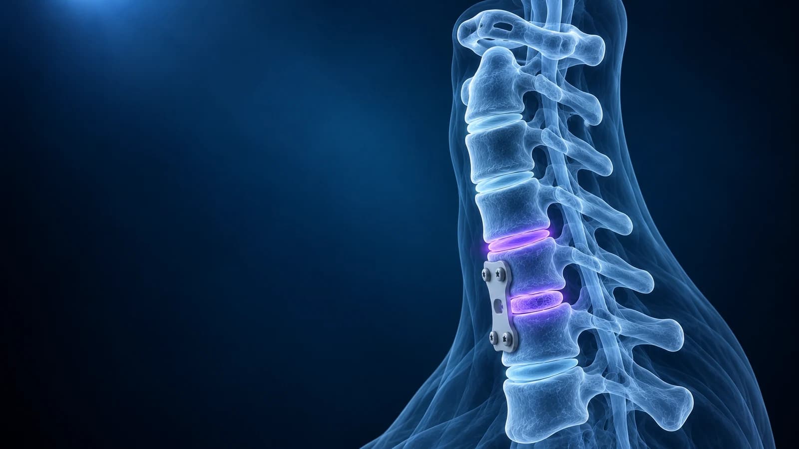
Yes, a neck MRI can usually be done and read after a cervical fusion.
Modern titanium plates, screws and spacers create a dark smudge of “susceptibility artifact” around the fused level, but the discs above and below, the spinal cord and the nerve exits are normally still visible. MRI after fusion mainly looks for new pressure on the cord or nerves, especially at the neighbouring levels. Whether the bones have actually fused and whether the hardware is secure is judged better on X-ray and CT.
This guide is for people who had an anterior cervical discectomy and fusion (ACDF) or a similar neck operation and now have a new scan, new symptoms or a report full of post-surgical wording. It explains what each test can show, the phrases you are likely to read and the symptoms that should not wait. It is educational; your surgeon remains the best person to compare the images with how you are doing.
Is it safe to have an MRI with a plate and screws in my neck?
For most modern spinal implants, yes, under the conditions set by the manufacturer. Plates, screws and cages used for neck fusion are typically titanium or plastic-like polymers, which are not pulled by the magnet. Tell the MRI team about every operation and bring your implant card or operation note if you have one, because a few devices, such as some artificial discs or stimulators, need specific settings. The scanning team, not the internet, makes the final safety check.
What does “hardware artifact limits evaluation” mean?
Metal distorts the magnetic field nearby, so the area right around the screws and plate looks dark or warped, sometimes with a bright rim. The radiologist is telling you that small details at the fused level, such as a sliver of remaining disc behind the spacer, cannot be judged confidently. Titanium causes far less artifact than stainless steel, and many scanners offer metal artifact reduction sequences that shrink the smudge further. When the fused level itself is the question, CT is usually the better tool; the American College of Radiology notes that CT can complement radiographs for new or changing symptoms after neck surgery.
What is adjacent segment disease after a neck fusion?
Once one level no longer moves, the levels above and below share more of the bending. Over time they can develop the same wear that led to the first operation: disc bulging, a disc osteophyte complex, facet joint enlargement and narrowing of the nerve exits. Reports call the imaging change “adjacent segment degeneration”. When it causes new symptoms, such as arm pain from a pinched root at the new level (cervical radiculopathy) or signs of cord compression (cervical myelopathy), surgeons call it adjacent segment disease.
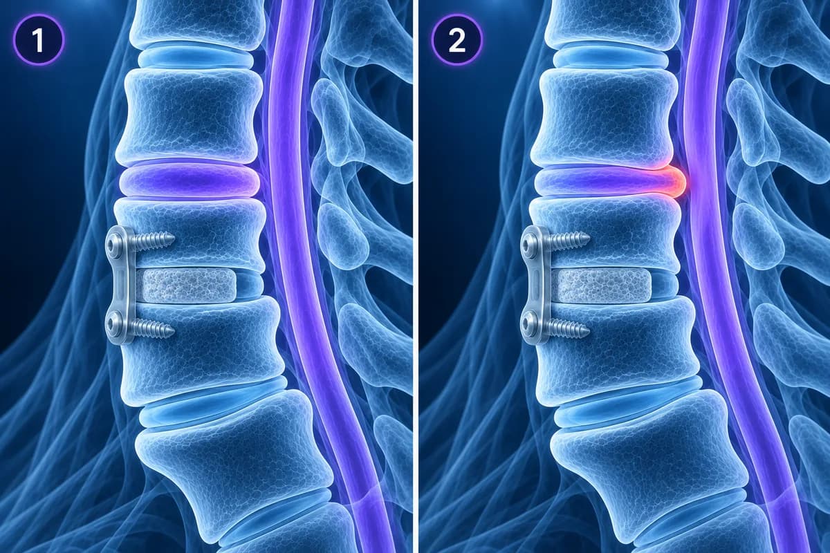
A classic long-term study of 374 patients followed after anterior fusion found new symptomatic disease at a neighbouring level at a fairly steady rate of 2.9% per year, predicting that about a quarter of patients would be affected within ten years. The risk was highest after single-level fusions involving C5 or C6 and when the neighbouring discs already looked worn before surgery, and the authors considered it mostly the natural progression of spondylosis. A 2024 meta-analysis of radiographic risk factors linked adjacent segment disease to less lordosis after surgery and to a canal that was narrow from birth. Wear at a neighbouring level on a scan is common and does not by itself mean another operation is needed.
Which post-surgery phrases might appear on my report?
- “Post-surgical changes of ACDF at C5–C6 with anterior plate, screws and interbody spacer”: a description of the operation that was done, not a problem.
- “Hardware intact” or “no evidence of hardware failure”: no broken or backed-out screws are seen, a statement made more confidently on X-ray or CT.
- “Lucency around the screws”: a dark halo in the bone around a screw on X-ray or CT, which can mean the screw is loosening or the level has not fused.
- “Residual” or “recurrent foraminal stenosis”: a nerve exit at the operated or a nearby level is still, or again, narrowed.
- “Myelomalacia” or “persistent T2 hyperintense cord signal”: a scar-like change in the cord from compression before surgery. It often remains after a successful decompression and is judged by comparing with earlier scans.
- “Prevertebral soft tissue swelling” or “small post-operative fluid collection”: expected in the first weeks after surgery from the front, important only when it is large or growing.
For wording that is not specific to surgery, use cervical MRI report phrases explained.
How do doctors check whether a neck fusion has healed?
Not with MRI. Bone needs months to bridge across the spacer, and solid fusion is judged on standing X-rays and CT. On flexion-extension X-rays, a fused level should not move between the bent-forward and tilted-back views. On CT, the report looks for “bridging trabecular bone” through or around the spacer. A level that keeps moving with a gap or halo around the implant is called a “pseudarthrosis” or non-union; it does not always cause symptoms, but it can explain persistent pain or a loosening screw. Our comparison of CT and MRI for the neck explains why bone questions favour CT.
When is contrast added to an MRI after neck surgery?
Gadolinium contrast is mostly used when infection is a concern, or when the team needs to tell scar tissue from a recurrent disc fragment. Scar enhances fairly evenly, while a disc fragment usually does not, apart from a thin rim. Infection after spine surgery can involve the disc space and neighbouring bone, which is covered on our discitis and osteomyelitis page. A routine follow-up scan for arm symptoms usually does not need contrast.
Which symptoms after a neck fusion need urgent care?
In the days after surgery from the front, growing neck swelling with difficulty swallowing or breathing is an emergency. At any stage, seek prompt care for fever, wound redness or discharge, new or worsening weakness or numbness in the arms or hands, clumsy hands, a change in balance or walking, new bladder or bowel problems, or a sudden increase in pain after a fall. Do not wait for a scheduled scan if any of these appear; contact your surgical team or emergency services.
How can I compare my scans before and after surgery?
Open the old and new MRI in our free MRI viewer, or a post-operative CT in the free CT viewer; files stay in your browser. Line up the same sagittal T2 slice on both, count down from C2 to the fused level, and then look one level up and one level down. Seeing whether the fluid around the cord has opened up at the operated level, and whether a neighbouring level has changed, makes the surgeon’s explanation much easier to follow. The basics of orientation are in how to read a cervical spine MRI.
Frequently Asked Questions
Can an MRI see through the metal in my neck?
Partly. The metal blurs a small area around the plate and screws, but the cord and the levels above and below are usually clear. If the fused level itself needs checking, CT is often added.
Why does my cord still show a bright spot after surgery?
Surgery removes the pressure but does not erase changes already present in the cord. A bright spot or myelomalacia often persists. What matters is whether it is new or larger compared with earlier scans and how your symptoms are changing.
Does adjacent segment degeneration mean I need another operation?
Not necessarily. Many people have wear next to a fusion without symptoms, and symptomatic cases often respond to non-surgical care first. Further surgery is considered for persistent or worsening nerve or cord symptoms that match the imaging.
How soon after neck fusion can I have an MRI?
Whenever it is clinically needed. Early scans show expected post-operative swelling that can be hard to separate from problems, so routine imaging is usually timed by your surgeon, while urgent symptoms are scanned without delay.
Can AI explain my post-fusion MRI report?
AI can translate post-surgical terms and point to the levels they describe, which helps you prepare for a follow-up visit. It is not a radiologist or surgeon and cannot judge your images against your operation and examination.
Sources
- Hilibrand AS, Carlson GD, Palumbo MA, et al. Radiculopathy and myelopathy at segments adjacent to the site of a previous anterior cervical arthrodesis. J Bone Joint Surg Am. 1999;81(4):519-528.
- Mesregah MK, Baker M, Yoon C, et al. Radiographic risk factors for adjacent segment disease following anterior cervical discectomy and fusion (ACDF): a systematic review and meta-analysis. Global Spine J. 2024;14(7):2183-2200.
- Expert Panel on Neurological Imaging; McDonald MA, Kirsch CFE, et al. ACR Appropriateness Criteria Cervical Neck Pain or Cervical Radiculopathy. J Am Coll Radiol. 2019;16(5S):S57-S76.
Related Articles
Why flexion and extension neck X-rays are ordered, how the test is done safely, which measurements suggest instability, why they are not the first test after an injury, and what the report wording means.
Why C5-C6 and C6-C7 are the neck discs that herniate most, which nerve root each one pinches, how the arm symptoms differ, and what protrusion, extrusion and foraminal mean on your MRI.
A plain-language guide to your neck MRI: how to count from C2 down to C7, which slices show the cord and the nerve exits, and which artifacts make a cervical scan look worse than it is.
Related Conditions
Understand your imaging before your next appointment
MRI, X-ray or CT from a CD, folder or ZIP. A PDF report or photos of the films work too.
Free to open and preview, no account needed. An optional AI report from $10 explains your images in plain language, with questions for your doctor.
Your raw files stay on this device. Only rendered images and minimal context are sent when you request an AI report.
Medical Disclaimer: This page is for informational and educational purposes only. It does not constitute medical advice, diagnosis, or treatment. AI-generated analysis may contain errors. Always consult a qualified healthcare professional for medical decisions. Full Disclaimer