
Neck X-ray vs CT vs MRI: Which Test Comes First?
Which neck scan comes first: X-ray or none for routine pain, MRI for a pinched nerve or cord signs, CT after injury under the NEXUS and Canadian C-spine rules.
Key Takeaways
- Ordinary neck pain without warning signs often needs no scan; an X-ray is the usual first picture
- MRI without contrast is the test for persistent pinched-nerve symptoms and signs of cord pressure
- After an injury, NEXUS or the Canadian C-spine rule decides whether to image, and CT is the first test for adults who need it
- X-ray and CT see bone; only MRI shows discs, the spinal cord and nerve roots directly
- A cervical X-ray is about 0.2 mSv and a spine CT about 6 mSv on average; MRI uses no radiation
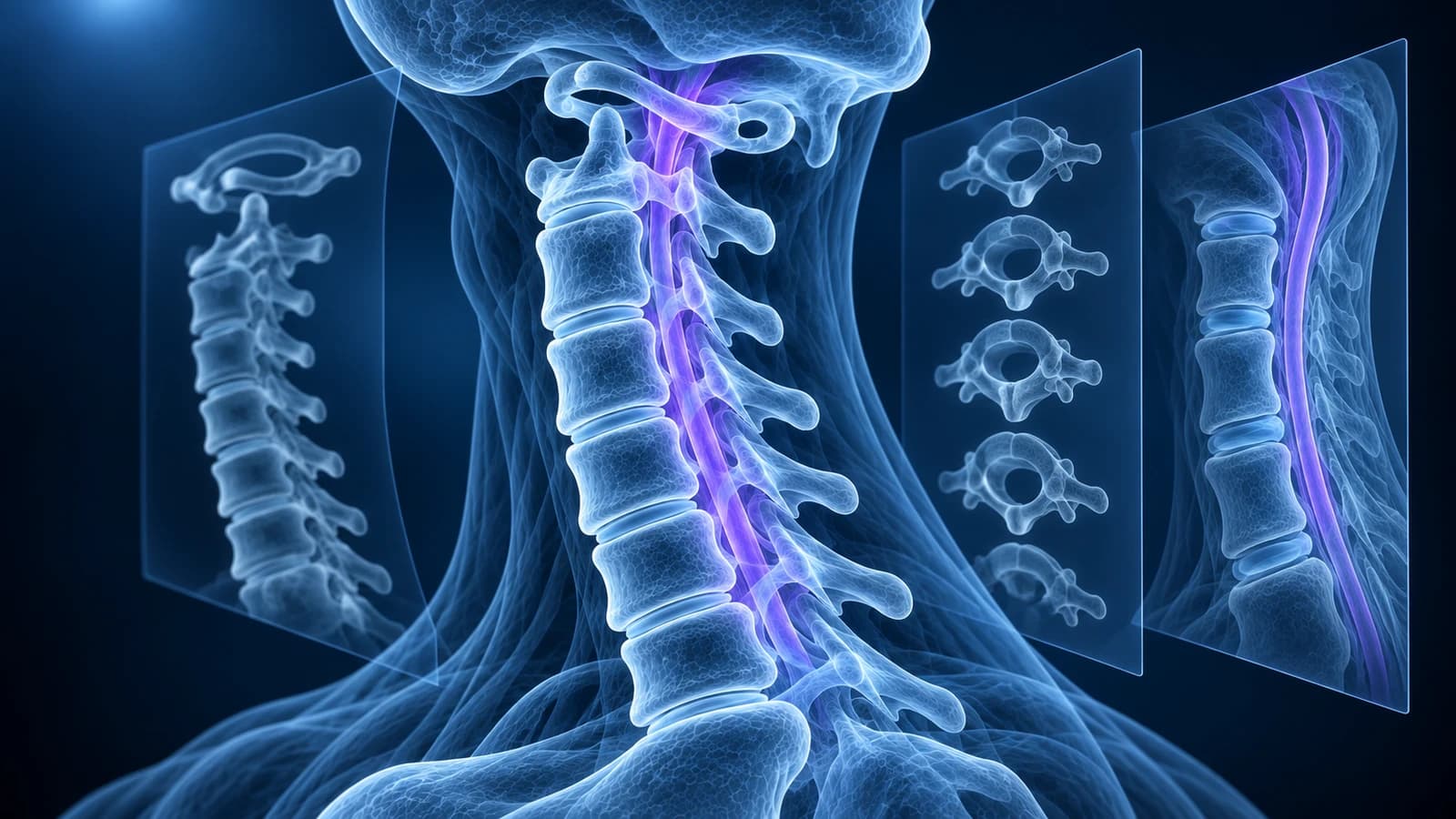
For everyday neck pain without arm symptoms, injury or warning signs, the first step is usually no imaging at all, or a plain X-ray. For arm pain from a pinched nerve that persists, or for signs of spinal cord pressure, MRI without contrast is the test. After an injury, an adult who meets the NEXUS or Canadian C-spine criteria for imaging gets a CT first.
The three tests are not a ladder from cheap to best. An X-ray, a CT and an MRI each see different tissues, and a result that is normal on one can be abnormal on another. This guide matches each common neck question to the test that answers it, explains what each scan shows and misses, and puts the radiation of X-ray and CT into plain numbers. It is general education based on published imaging guidelines, not a diagnosis or a referral.
When is the choice of scan an emergency decision?
Do not wait for a booked scan if neck pain comes with any of the following. Go to an emergency department or call emergency services, and keep the neck still after a serious injury until you are assessed.
- A road collision, a fall from height, a diving accident or a blow to the head, especially at 65 or older
- Numbness, tingling or weakness in the arms or legs after an injury
- Clumsy hands, a new unsteady walk, or new trouble controlling the bladder or bowel
- Fever, chills or feeling very unwell with neck pain
- A history of cancer with new, constant neck pain that is worse at night
Which test comes first for neck pain without an injury?
The American College of Radiology Appropriateness Criteria for cervical pain or cervical radiculopathy, updated in 2024, sort patients by symptoms rather than by preference. For new neck pain with no arm symptoms, no trauma and no red flags, an X-ray may be appropriate, while MRI and CT are rated usually not appropriate as a first test. For many people the honest answer is that no scan is needed yet.
Once arm pain, tingling or weakness follows a nerve-root pattern, MRI moves forward. For recent arm symptoms the criteria rate both an X-ray and an MRI without contrast as may be appropriate, and for arm symptoms that have lasted, MRI without contrast becomes usually appropriate. That is the typical path for cervical radiculopathy, where the question is which disc or spur is touching which nerve root.
Red flags change the order again. For suspected infection or a known cancer, the criteria rate MRI without and with contrast as usually appropriate and plain X-ray as usually not appropriate, because the early changes sit in bone marrow and soft tissue. After previous neck surgery, X-ray, CT and MRI are all rated usually appropriate: X-ray checks the hardware and alignment, CT checks whether a fusion has healed, and MRI looks for new pressure on nerves.
Which test comes first after a neck injury?
After trauma the first question is not which scan but whether any scan is needed, and two validated decision rules answer it. The NEXUS low-risk criteria allow a clinician to skip imaging when the person has no tenderness over the midline of the back of the neck, is not intoxicated, is fully alert, has no neurological deficit and has no other painful injury that could distract from neck pain.
The Canadian C-spine rule, built for alert and stable adults, sends anyone aged 65 or over, anyone with a dangerous mechanism of injury and anyone with tingling in the limbs straight to imaging. Others who have a low-risk feature, such as a simple rear-end collision or being able to sit up and walk, are asked to turn the head 45 degrees each way; if they can, no imaging is needed. In a head-to-head study of 8,283 alert, stable trauma patients, the Canadian rule picked up 99.4% of important neck injuries against 90.7% for NEXUS, and would have meant fewer X-rays.
When an adult does meet the criteria for imaging, the ACR criteria for acute spinal trauma make CT without contrast the first test, because it is fast, accurate and available around the clock, and they rate CT and MRI as usually not appropriate for low-risk adults the rules have cleared. MRI is added when there are neurological symptoms, when CT is normal but a ligament or disc injury is suspected, or when the cord needs to be seen. Our page on cervical spine fracture covers what happens next, and what a whiplash MRI shows covers the soft-tissue side of lower-energy injuries.
What does each neck scan show, and what does it miss?
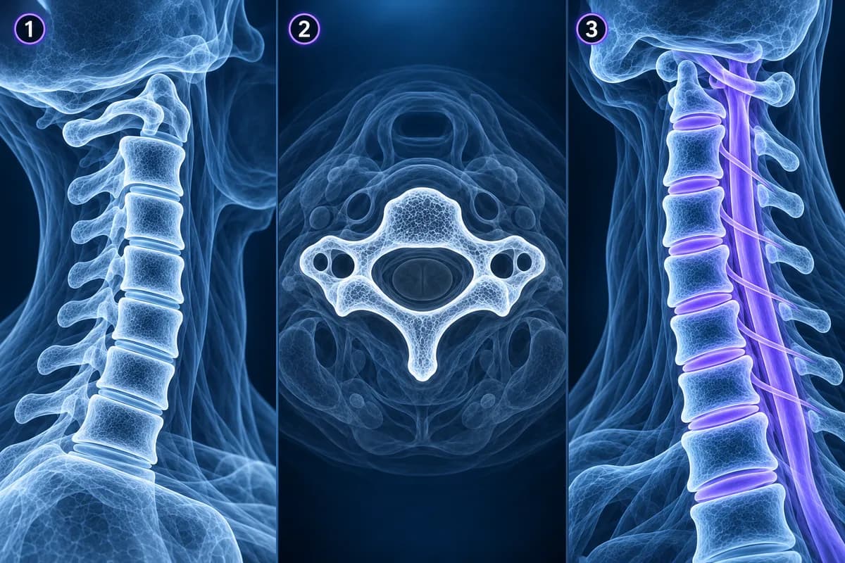
An X-ray passes a small dose of radiation through the neck and records the shadow of the bones. It shows alignment, the height of each disc space, bone spurs and the shape of the vertebrae in seconds, and bending views can reveal a slip that only appears with movement. It cannot show the discs themselves, the nerves or the spinal cord, and it misses many fractures, which is why it has largely been replaced by CT after significant trauma. The cervical spine X-ray guide walks through each view.
CT rotates an X-ray beam around the neck and rebuilds the bones as thin slices that can be viewed from any angle. It is the best test for fractures, for the bony edges of the nerve openings, for checking whether a fusion has healed, and for calcified ligaments such as OPLL. It shows discs and the cord only faintly, so a soft disc herniation or swelling inside the cord can be missed. With dye injected into the spinal fluid, a CT myelogram outlines the cord and nerve roots for people who cannot have an MRI.
MRI uses a magnetic field and radio waves instead of radiation. It is the only one of the three that shows the discs, the spinal cord, the nerve roots, the ligaments and the bone marrow directly, which is why it answers questions about pinched nerves, cord compression, infection and tumours. It is less sharp than CT for fine bone detail, takes longer, is noisy, and is not possible with some implanted devices.
Which neck scan is used for which question?
- Ordinary neck pain without arm symptoms or warning signs: often no imaging; an X-ray if a first picture is wanted
- Arm pain, tingling or weakness from a pinched nerve that persists: MRI without contrast, with CT added when a bony spur needs to be measured
- Clumsy hands, balance change or other signs of cord pressure: MRI without contrast, promptly
- Injury that meets NEXUS or Canadian C-spine imaging criteria: CT first, MRI if there are neurological signs or a suspected ligament injury
- Suspected instability at a known slip or after surgery: X-rays in flexion and extension to show movement
- Suspected infection or cancer spread: MRI without and with contrast
- Unable to have an MRI: CT, or a CT myelogram when the nerves and cord must be seen
For the most common follow-up dilemma, a pinched nerve where both CT and MRI are on the table, see CT vs MRI for cervical radiculopathy. Movement views are explained in cervical flexion-extension X-rays, and signs of cord pressure are covered in cervical myelopathy.
How much radiation is a neck X-ray or CT scan?
Radiation dose is measured in millisieverts (mSv). Everyone receives about 3 mSv a year from natural background radiation. A widely used radiology catalogue puts an average cervical spine X-ray at about 0.2 mSv, roughly a few weeks of background, and an average CT of the spine at about 6 mSv, roughly two years of background. Actual doses vary with the scanner, the number of views and your build.
MRI uses no ionising radiation at all. That does not make it the safer choice in every case: after a serious injury, a fast CT that finds a fracture matters far more than the dose, and the risk from a single scan is small. Where radiation does deserve thought is in children and in people having repeated scans, and the thyroid gland sits in the front of the neck, so ask whether a test will change your care if you have had several CTs before.
How can you look at your own neck X-ray, CT or MRI?
Ask the imaging department for the DICOM files as well as the written report. Alignment, spurs and fractures are easiest to follow in the free X-ray viewer or the free CT viewer, where you can scroll slices and adjust the bone window. Discs, the cord and the nerve roots belong in the free MRI viewer. All three open files in your browser without uploading them. For the report wording itself, cervical MRI report phrases explained translates the common terms.
Frequently Asked Questions
Is an MRI better than an X-ray for neck pain?
Better at some questions, worse at others. MRI shows discs, nerves and the cord, but it also finds age-related changes in most adults that may have nothing to do with the pain. For uncomplicated neck pain, guidelines do not recommend MRI first; an X-ray, or no imaging, is the usual start.
Why did the emergency department do a CT and not an X-ray of my neck?
Because CT finds neck fractures much more reliably than plain X-rays and is quick to perform. The ACR criteria for acute spinal trauma make CT the first test for adults whose injury meets the NEXUS or Canadian C-spine criteria for imaging.
My neck CT was normal. Why do I need an MRI as well?
A normal CT rules out most fractures, but it shows ligaments, discs and the spinal cord poorly. If you have numbness, weakness or pain that suggests a soft-tissue injury, MRI is used to look at the structures CT cannot see.
Can an X-ray show a herniated disc in the neck?
No. An X-ray can show a narrowed disc space or spurs that hint at disc wear, but the disc itself and any herniation are invisible. MRI shows the disc and whether it is pressing on a nerve root, as described in cervical disc herniation.
Which neck scan is cheapest?
An X-ray is usually the least expensive, followed by CT, with MRI typically the most expensive. The right test is the one that answers your clinical question, and our guide to cervical MRI cost explains what sets the price of an MRI.
Sources
- Expert Panel on Neurological Imaging; Eldaya RW, Parsons MS, Hutchins TA, et al. ACR Appropriateness Criteria Cervical Pain or Cervical Radiculopathy: 2024 Update. J Am Coll Radiol. 2025;22(5S):S136-S162.
- Expert Panel on Neurological Imaging; Hassankhani A, Freeman CW, Banks J, et al. ACR Appropriateness Criteria Acute Spinal Trauma: 2024 Update. J Am Coll Radiol. 2025;22(5S):S48-S66.
- Hoffman JR, Mower WR, Wolfson AB, et al. Validity of a set of clinical criteria to rule out injury to the cervical spine in patients with blunt trauma. N Engl J Med. 2000;343(2):94-99.
- Stiell IG, Wells GA, Vandemheen KL, et al. The Canadian C-spine rule for radiography in alert and stable trauma patients. JAMA. 2001;286(15):1841-1848.
- Stiell IG, Clement CM, McKnight RD, et al. The Canadian C-spine rule versus the NEXUS low-risk criteria in patients with trauma. N Engl J Med. 2003;349(26):2510-2518.
- Mettler FA Jr, Huda W, Yoshizumi TT, Mahesh M. Effective doses in radiology and diagnostic nuclear medicine: a catalog. Radiology. 2008;248(1):254-263.
Related Articles
MRI is usually the first scan for a pinched nerve in the neck, while CT shows bone spurs, OPLL and foraminal narrowing in finer detail. Learn when each is used, when CT myelography is needed, and what the report phrases mean.
What a neck X-ray can and cannot show: the standard views, the four alignment lines, disc space narrowing, bone spurs and the Torg-Pavlov ratio, and when CT or MRI is needed instead.
Most neck pain does not need an MRI. Learn the warning signs that do call for imaging, why X-ray or CT comes first after an injury, and how to read the age-related findings a neck MRI almost always shows.
Related Conditions
Understand your imaging before your next appointment
MRI, X-ray or CT from a CD, folder or ZIP. A PDF report or photos of the films work too.
Free to open and preview, no account needed. An optional AI report from $10 explains your images in plain language, with questions for your doctor.
Your raw files stay on this device. Only rendered images and minimal context are sent when you request an AI report.
Medical Disclaimer: This page is for informational and educational purposes only. It does not constitute medical advice, diagnosis, or treatment. AI-generated analysis may contain errors. Always consult a qualified healthcare professional for medical decisions. Full Disclaimer