
CT vs MRI for Cervical Radiculopathy: Which Scan Shows a Pinched Nerve?
MRI is usually the first scan for a pinched nerve in the neck, while CT shows bone spurs, OPLL and foraminal narrowing in finer detail. Learn when each is used, when CT myelography is needed, and what the report phrases mean.
Key Takeaways
- MRI without contrast is the usual first scan when a pinched nerve in the neck needs imaging
- CT is better for bone spurs, calcified discs, OPLL, fractures and hardware
- CT myelography is reserved for people who cannot have MRI or when MRI leaves the question open
- Surgeons often review both, because whether compression is soft disc or bone changes the approach
- Progressive weakness, symptoms in both arms or the legs, clumsy hands or bladder change need prompt care first
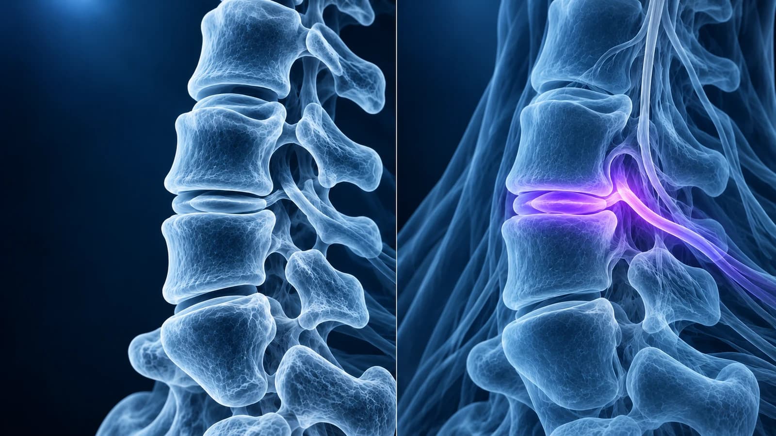
For cervical radiculopathy, a pinched nerve root in the neck, MRI without contrast is usually the first scan when imaging is needed, because it shows the disc, the nerve root and the spinal cord directly. CT is better at bone: it shows the spurs that narrow a nerve opening, ossified ligament and fractures in finer detail, and it is the alternative when MRI is not possible.
The choice matters because a nerve root in the neck can be squeezed by two quite different things. A “soft” disc herniation is common in younger adults and is best seen on MRI. A “hard” narrowing made of bone spurs from the small joints at the side of the disc, called uncovertebral joints, is more common after middle age and is best outlined on CT. This page compares the two tests for this one question; it is educational, and the test that suits you is a decision for your clinician.
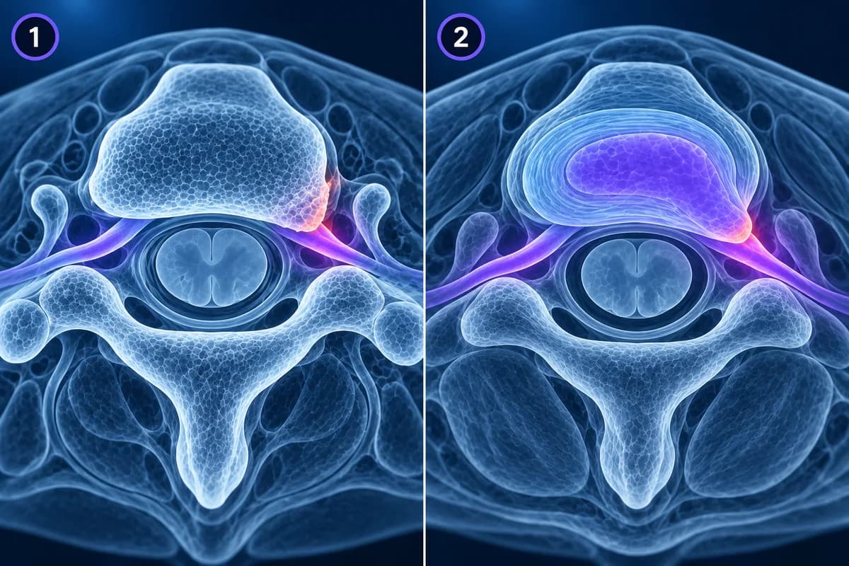
When is arm pain from the neck urgent?
The choice of scan comes after safety. Seek prompt medical care if arm weakness is getting worse, if symptoms involve both arms or the legs, if your hands become clumsy or your walking unsteady, if bladder or bowel control changes, or if the pain follows significant trauma or comes with fever, a cancer history or weakened immunity. Those features suggest more than a single pinched nerve and change the urgency of imaging.
Which scan do guidelines recommend for a pinched nerve in the neck?
Most pinched nerves in the neck improve with time and non-operative care, so many people never need a scan. The ACR Appropriateness Criteria for cervical neck pain or radiculopathy reserve advanced imaging for radiculopathy that persists or progresses despite treatment, for red flags, and for planning an injection or operation. In those situations MRI of the cervical spine without contrast is the preferred study. CT and CT myelography are listed as options mainly for people who cannot have MRI or when MRI does not answer the question.
Whichever test is used, the finding has to match the arm. In a large MRI study of adults with no neck symptoms, 87.6% had disc bulging, so a bulge on the wrong side or at the wrong level does not explain a pinched nerve. In practice, many surgeons look at both scans before operating. MRI shows what is pressing on the root and whether the cord is also involved; CT shows how much of that pressure is bone, which changes how the surgeon approaches the level. For the broader question of whether you need a scan at all, see do you need an MRI for neck pain.
What does MRI show that CT cannot?
- The nerve root itself, surrounded by bright fluid, and the point where it is displaced or flattened
- Soft disc material: a protrusion or extruded fragment pushing into the nerve opening, which on CT can blend into the surrounding tissue
- The spinal cord, including flattening and T2 hyperintense cord signal, which turns a nerve-root question into a cord question
- Disc hydration, annular fissures and bone marrow changes next to a worn disc
- Infection, tumour and inflammation, especially with contrast, when a red flag is present
MRI uses no ionising radiation. Its drawbacks are time, usually around half an hour lying still, noise, difficulty for people with claustrophobia, and restrictions for some implanted devices. Metal from earlier neck surgery can blur the images near the hardware, a limitation discussed in cervical MRI after fusion.
What does CT show better than MRI?
- Uncovertebral and facet spurs, measured precisely as they project into the nerve opening
- Calcified disc material and ossified ligament, which MRI can show only as dark tissue that is hard to tell apart from a soft disc
- Ossification of the posterior longitudinal ligament, a bony plate behind the vertebral bodies described in OPLL
- Fractures, fusion status after surgery, and the position of screws and plates
CT takes seconds and works for nearly everyone, including people with pacemakers or severe claustrophobia. Its main costs are radiation, with the thyroid gland lying in the scanned area, and the fact that a plain CT shows the nerve and disc only faintly. Oblique reconstructions angled along each nerve opening make foraminal narrowing much easier to judge than standard straight slices.
When is a CT myelogram used instead?
A CT myelogram adds contrast injected into the fluid around the spinal cord through a lumbar puncture, then a CT scan of the neck. The contrast outlines the cord and each nerve root sleeve, so a root that does not fill with contrast is being compressed. It combines the bone detail of CT with an indirect picture of the nerves.
Because it is invasive, it is kept for specific cases: people who cannot have MRI because of a device that is not MRI-conditional, people with metal from earlier surgery that obscures the MRI, and situations where MRI and the examination disagree and a surgeon needs a clearer answer before operating. Possible side effects include a headache after the lumbar puncture.
What do radiculopathy report phrases mean on CT and MRI?
- “Uncovertebral hypertrophy” or “uncovertebral joint arthropathy” — spurs on the small side joints of the vertebra, the most common bony cause of a pinched nerve in the neck
- “Mild, moderate or severe foraminal narrowing” — how much of the nerve opening is lost; see cervical foraminal stenosis
- “Foraminal disc protrusion contacting the exiting C7 nerve root” — soft disc material in the opening, touching the named nerve
- “Disc osteophyte complex” — disc and spur fused into one ridge, a term used on both tests that does not say how much is bone; CT answers that
- “Facet arthropathy” — wear of the joints at the back, which can narrow the opening from behind
- “Calcified” or “ossified” — on a CT report, material that is hard, which helps a surgeon plan
In the neck, a nerve root exits above the vertebra it is named after, so the C6 root leaves at C5–C6 and the C7 root at C6–C7. Those two levels account for most cervical radiculopathy, as explained in C5–C6 and C6–C7 disc herniation explained. A C6 root problem classically affects the thumb side of the hand, a C7 root problem the middle finger and the back of the arm, which is how a clinician checks whether the side and level on the scan fit your symptoms.
Can you compare your own CT and MRI side by side?
If you have both studies on a disc or download, open them at the same level: find the disc named in the report on the sagittal series, then switch to the axial slice through it. On MRI, look for the dark nerve root in the bright fluid on each side; on CT, look at the shape of the bony opening and any spur pointing into it. The free MRI viewer and free CT viewer open DICOM files locally in your browser. For orientation on the MRI series, see how to read a cervical spine MRI, and for the condition itself, cervical radiculopathy and cervical disc herniation.
Frequently Asked Questions
Can a CT scan miss a herniated disc in the neck?
Yes. Soft disc material and nerve roots have similar density on CT, so a small or foraminal herniation can be hard to see, especially in a slim neck with little fat around the nerves. CT is more reliable when the compression is bony. When a soft disc is suspected and the CT is unclear, MRI is the usual next step.
Why did my doctor order a CT after my MRI?
Usually to see how much of the narrowing is bone. MRI can show a dark ridge without saying whether it is calcified disc, spur or ossified ligament. Before surgery, CT also maps the anatomy for screws and implants. Ordering a CT after an MRI does not by itself mean something worse was found.
How much radiation is a cervical spine CT?
A neck CT involves more radiation than a set of neck X-rays, and the thyroid lies in the scanned area. The dose is considered acceptable when the scan is needed to answer a clinical question, but it is one reason MRI is preferred for nerve problems when it is available. Ask the imaging department about dose if you have had many scans.
Is an MRI with contrast better for a pinched nerve?
Not usually. Contrast does not improve the view of a disc pressing on a nerve root in a neck that has not had surgery. It is added when infection, a tumour or inflammation is suspected, or to separate scar tissue from a new disc herniation after an operation.
Sources
- Expert Panel on Neurological Imaging; McDonald MA, Kirsch CFE, et al. ACR Appropriateness Criteria Cervical Neck Pain or Cervical Radiculopathy. J Am Coll Radiol. 2019;16(5S):S57-S76.
- Iyer S, Kim HJ. Cervical radiculopathy. Curr Rev Musculoskelet Med. 2016;9(3):272-280.
- Nakashima H, Yukawa Y, Suda K, et al. Abnormal findings on magnetic resonance images of the cervical spines in 1211 asymptomatic subjects. Spine (Phila Pa 1976). 2015;40(6):392-398.
Related Articles
A plain-language guide to "disc osteophyte complex" on a cervical MRI: what the ridge is made of, how it differs from a herniated disc, and what effacing the thecal sac, cord contact and cord compression mean.
Why C5-C6 and C6-C7 are the neck discs that herniate most, which nerve root each one pinches, how the arm symptoms differ, and what protrusion, extrusion and foraminal mean on your MRI.
A plain-language guide to your neck MRI: how to count from C2 down to C7, which slices show the cord and the nerve exits, and which artifacts make a cervical scan look worse than it is.
Related Conditions
Understand your imaging before your next appointment
MRI, X-ray or CT from a CD, folder or ZIP. A PDF report or photos of the films work too.
Free to open and preview, no account needed. An optional AI report from $10 explains your images in plain language, with questions for your doctor.
Your raw files stay on this device. Only rendered images and minimal context are sent when you request an AI report.
Medical Disclaimer: This page is for informational and educational purposes only. It does not constitute medical advice, diagnosis, or treatment. AI-generated analysis may contain errors. Always consult a qualified healthcare professional for medical decisions. Full Disclaimer