
Cervical Spinal Stenosis: What Central Canal Narrowing on Your Neck MRI Means
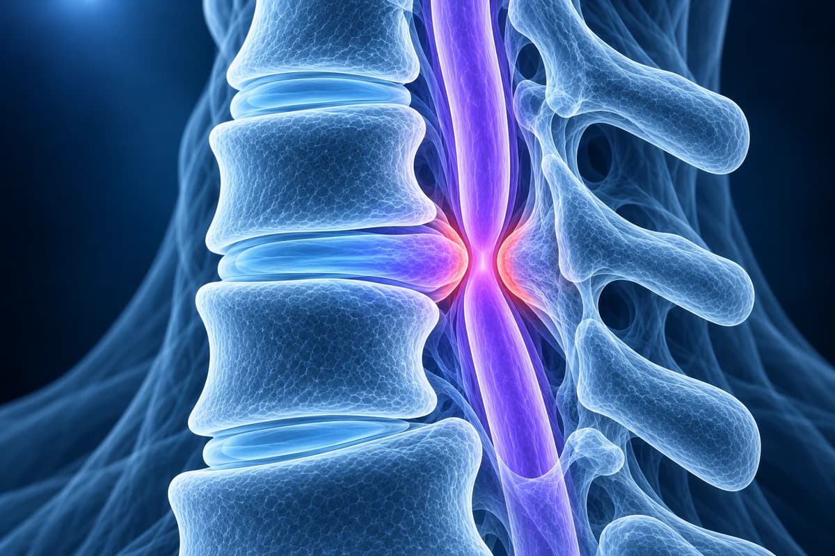
Cervical spinal stenosis means the central canal that carries the spinal cord through the neck is narrower than normal, usually because of disc, bone-spur and ligament changes. A bony front-to-back canal diameter below about 13 mm is considered narrow. On MRI, what matters most is whether the narrowing only pushes fluid aside, touches the cord, or actually compresses it.
Common Symptoms
- Often no symptoms at all: many narrow canals are found on a scan done for something else
- Neck stiffness and aching that is worse when looking up for a long time
- Tingling or numbness in both hands rather than in one finger pattern
- Fingers that feel less nimble with buttons, writing or typing
- A brief electric feeling down the back or limbs when the chin drops to the chest
- Legs that feel heavy or less steady, especially in the dark or on uneven ground
When does a narrow neck canal need urgent attention?
Seek emergency care if you develop weakness in both arms or both legs, sudden difficulty walking, new loss of bladder or bowel control, or numbness spreading across the chest or trunk. A narrow canal leaves the cord little spare room, so after a fall, a car crash or a hard tackle, even without a fracture, new tingling or weakness in the limbs needs same-day assessment. Tell the emergency team that a scan has shown cervical stenosis.
Book a prompt, non-emergency appointment if your hands have been getting clumsier, your balance has slowly changed, or you feel the electric feeling when bending your head forward. Neck pain with fever, unexplained weight loss, a past cancer or a weakened immune system also needs urgent review, because infection or a tumour can narrow the canal too. This page explains imaging wording; it is not a diagnosis.
Why does the central canal in the neck become narrow?
The central canal is a tunnel formed by the back of the vertebral bodies and discs in front and the bony arch (laminae) and ligamentum flavum behind. Narrowing is either acquired, built up over years of wear, or congenital, meaning the tunnel was small from the start. In real life the two often add together: a person with a borderline canal reaches the point of cord contact with less wear than someone with a roomy one.
Acquired narrowing usually has more than one ingredient at the same level. A worn disc and the bone spurs along its rim form a disc-osteophyte complex that bulges backward. The ligamentum flavum thickens and folds inward when the disc loses height, pinching the canal from behind. A vertebra may slip slightly forward on the one below. In some people the ligament on the back of the vertebral bodies turns to bone, called OPLL, which can narrow the canal across several levels. Together these are part of cervical spondylosis.
Congenital or developmental stenosis comes from short pedicles, the bony struts that set the depth of the canal. In a study of 1,066 adult skeletons, a sagittal canal diameter under 13 mm was strongly linked with a congenitally narrow canal. Birth differences such as Klippel-Feil syndrome and Down syndrome are also described as conditions that leave the cord more vulnerable to later degeneration.
What do mild, moderate and severe central canal stenosis mean?
There is no single scale that every radiologist uses, so the same canal can be called mild in one report and moderate in another. The words are most useful when you know what the radiologist saw on the sagittal T2 images. One widely cited MRI scheme, published by Kang and colleagues in 2011, sorts central stenosis into four grades based on how the fluid and the cord look:
- Grade 0: no stenosis. A rim of bright spinal fluid surrounds the cord.
- Grade 1: more than half of the fluid space in front of or behind the cord is obliterated, but the cord keeps its shape.
- Grade 2: the cord itself is deformed, flattened or indented, without a signal change inside it.
- Grade 3: the cord is compressed and shows a bright signal change on T2-weighted images.
In that scheme, grades 0 to 1 are treated as less significant and grades 2 to 3 as significant stenosis. When a report says only "moderate" or "severe", ask which of these it is closest to: fluid narrowed, cord touched, cord deformed, or cord deformed with signal change. That one question tells you far more than the adjective.
What does "central canal stenosis" wording mean on your MRI report?
- "Effacement of the ventral CSF" or "effacement of the thecal sac"
- The fluid in front of the cord has been squeezed away at that level, but the cord has not been reached. Ask whether any fluid remains behind the cord and whether the narrowing changes when you bend the neck.
- "Abuts the cord" or "contacts the ventral cord"
- The disc or spur now touches the cord surface without changing its shape. Ask whether this level fits your symptoms and what would prompt a repeat scan.
- "Indents", "flattens" or "deforms the cord"
- The cord has lost its normal oval outline, which is anatomical cord compression. Ask your clinician to check for signs of myelopathy, such as reflex changes, hand clumsiness and gait problems, because the next step depends on them.
- "Increased T2 cord signal" or "myelomalacia"
- A bright spot inside the compressed cord on T2 images, or softened cord tissue on longer-standing cases. This is the strongest imaging sign that the cord itself is affected, so ask for a spine specialist review rather than routine follow-up.
- "Ligamentum flavum buckling" or "ligamentum flavum hypertrophy"
- The ligament behind the cord is thick or folded and narrows the canal from behind. Ask whether the compression comes mainly from the front, from behind or both, because that guides any surgical approach.
- "Developmentally narrow canal" or "short pedicles"
- The canal was small before any wear began. Ask how much spare room is left and whether this changes your advice on contact sports or high-risk activities.
More phrases from neck reports are collected in cervical MRI report phrases explained.
Is central canal stenosis the same as foraminal stenosis or myelopathy?
No, the three terms describe different things. Central canal stenosis is about the main tunnel and the spinal cord inside it. Foraminal stenosis is narrowing of the small side openings where each nerve root leaves the spine; it tends to cause pain, tingling or weakness down one arm. A single report often lists both at the same level, because the same disc and spurs can encroach on the middle and the side of the canal at once.
Cervical myelopathy is not an imaging finding but a clinical diagnosis: the cord is not working normally, shown by symptoms and examination signs. Stenosis is the narrowed space; myelopathy is the cord damage that severe or long-standing stenosis can cause. Someone can have a tight canal on MRI without myelopathy, and the treatment conversation is very different for each.
Stenosis lower down in the back behaves differently again: the lumbar canal holds nerve roots rather than the cord, so it typically causes leg pain and heaviness when walking, covered on the lumbar spinal stenosis page.
Which scan shows cervical spinal stenosis best?
For ordinary neck pain without arm symptoms or warning signs, imaging is often not needed straight away, and plain X-rays are the usual first test when it is. The ACR Appropriateness Criteria for cervical pain or radiculopathy place MRI without contrast at the centre when symptoms persist, involve the arms, or come with red flags. When cord symptoms are suspected, the ACR criteria for myelopathy rate MRI without contrast as usually appropriate, because it is the only routine test that shows the cord, the fluid around it and any signal change.
X-rays cannot see the cord, but a side view lets the canal be measured against the vertebral body. In a 1986 study of athletes with transient cord symptoms, a canal-to-vertebral-body ratio under 0.80, now called the Torg-Pavlov ratio, marked significant stenosis. CT shows bone spurs and ossified ligament in fine detail and is often added before surgery. CT myelography is used when MRI is not possible. Contrast MRI is reserved for suspected infection, tumour or inflammation. After an injury, emergency teams use rules such as NEXUS or the Canadian C-spine Rule to decide on CT first, with MRI added when there are neurological symptoms.
For a guide to the sequences and views, see how to read a cervical spine MRI.
What we look for on cervical canal stenosis imaging
- Each level from C2–C3 to C7–T1, with whether fluid remains in front of and behind the cord
- Whether the cord is touched, indented or flattened, and over how many levels
- Bright T2 signal or dark T1 signal inside the cord at the narrowest point
- What does the narrowing: disc-osteophyte complex, buckled ligamentum flavum, OPLL or a slipped vertebra
- Signs of a developmentally narrow canal, such as short pedicles across many levels
- Accompanying foraminal narrowing at the same levels and the overall curve of the neck
Is a narrow neck canal dangerous if I have no symptoms?
Usually it is a finding to understand and monitor rather than to fix. The 2017 AO Spine guideline on degenerative cervical myelopathy does not recommend preventive surgery for people whose MRI shows cord compression without myelopathy. It recommends teaching them the warning symptoms and following them clinically, and it notes that people with compression and abnormal nerve tests or radiculopathy carry a higher risk of developing myelopathy.
The main practical risk is a sudden extension or flexion injury. In athletes, a narrow canal is linked with brief episodes of burning, numbness or weakness in all four limbs after a collision, called transient cord neurapraxia. A 1996 study of football players found that a Torg-Pavlov ratio of 0.80 or less was sensitive for these episodes but had a very low predictive value, so the ratio alone is not used to bar people from contact sport when the spine is stable. Any such episode, however, needs an MRI and specialist advice before returning to play.
How is cervical spinal stenosis treated?
Treatment follows symptoms, not the scan alone. Neck pain with a narrow canal but no cord signs is usually managed like other degenerative neck pain: staying active, physiotherapy focused on posture and strength, and short courses of pain relief. Your clinician may advise avoiding forceful neck manipulation and activities with a high risk of head-first impact.
When stenosis causes myelopathy, the AO Spine guideline recommends surgery for moderate and severe disease, and either surgery or supervised rehabilitation with close follow-up for mild disease. Surgery widens the canal from the front by removing disc and spur and fusing the level, or from behind by laminoplasty or laminectomy with fusion. If you have already had an operation, see cervical MRI after fusion for what later scans show.
Can I look at the narrow level on my own neck MRI?
Yes. Open your DICOM files privately in the free cervical MRI viewer, pick the sagittal T2 series, and scroll to the level your report names: look for whether the white fluid line in front of and behind the cord is still there. A neck CT opens in the free CT viewer. For a plain-language walkthrough of your images and report, upload them for an AI review. It explains findings, it does not diagnose, and new arm or leg symptoms should go to a clinician first.
Frequently Asked Questions
What is a normal cervical spinal canal diameter?
Canal size varies between people and levels, so there is no single cut-off. A front-to-back bony diameter below about 13 mm is strongly linked with a congenitally narrow canal, and on an X-ray a canal-to-vertebral-body ratio under 0.80 is a commonly used marker of stenosis.
Is moderate central canal stenosis serious?
It depends on what moderate means in your report. If fluid is narrowed but the cord keeps its shape and you have no cord symptoms, it is usually monitored. If the cord is flattened or shows signal change, or you have hand or walking problems, it needs specialist review.
What is the difference between spinal stenosis and cord compression?
Stenosis is any narrowing of the canal. Cord compression is the stage where the narrowing actually deforms the cord. Many people with stenosis on MRI have no cord compression at all.
Can cervical spinal stenosis get worse?
Degenerative narrowing tends to progress slowly over years, but many people never develop cord symptoms. A change in hand dexterity, balance or bladder control is the signal to get re-examined, rather than a fixed schedule of repeat scans.
Can I play contact sports with a narrow neck canal?
A narrow canal in a stable spine without symptoms does not automatically rule it out. An episode of burning, numbness or weakness in the limbs after a hit, cord signal change, or instability changes that advice, and the decision should be made with a spine or sports medicine specialist.
Sources
- Kang Y, Lee JW, Koh YH, et al. New MRI grading system for the cervical canal stenosis. AJR Am J Roentgenol. 2011;197(1):W134-W140.
- Bajwa NS, Toy JO, Young EY, Ahn NU. Establishment of parameters for congenital stenosis of the cervical spine: an anatomic descriptive analysis of 1,066 cadaveric specimens. Eur Spine J. 2012;21(12):2467-2474.
- Torg JS, Pavlov H, Genuario SE, et al. Neurapraxia of the cervical spinal cord with transient quadriplegia. J Bone Joint Surg Am. 1986;68(9):1354-1370.
- Torg JS, Naranja RJ Jr, Pavlov H, et al. The relationship of developmental narrowing of the cervical spinal canal to reversible and irreversible injury of the cervical spinal cord in football players. J Bone Joint Surg Am. 1996;78(9):1308-1314.
- Nouri A, Tetreault L, Singh A, Karadimas SK, Fehlings MG. Degenerative Cervical Myelopathy: Epidemiology, Genetics, and Pathogenesis. Spine (Phila Pa 1976). 2015;40(12):E675-E693.
- Expert Panel on Neurological Imaging; Eldaya RW, Parsons MS, Hutchins TA, et al. ACR Appropriateness Criteria Cervical Pain or Cervical Radiculopathy: 2024 Update. J Am Coll Radiol. 2025;22(5S):S136-S162.
- Expert Panel on Neurological Imaging; Agarwal V, Shah LM, Parsons MS, et al. ACR Appropriateness Criteria Myelopathy: 2021 Update. J Am Coll Radiol. 2021;18(5S):S73-S82.
- Fehlings MG, Tetreault LA, Riew KD, et al. A Clinical Practice Guideline for the Management of Patients With Degenerative Cervical Myelopathy: Recommendations for Patients With Mild, Moderate, and Severe Disease and Nonmyelopathic Patients With Evidence of Cord Compression. Global Spine J. 2017;7(3 Suppl):70S-83S.
Understand your imaging before your next appointment
MRI, X-ray or CT from a CD, folder or ZIP. A PDF report or photos of the films work too.
Free to open and preview, no account needed. An optional AI report from $10 explains your images in plain language, with questions for your doctor.
Your raw files stay on this device. Only rendered images and minimal context are sent when you request an AI report.
Medical Disclaimer: This page is for informational and educational purposes only. It does not constitute medical advice, diagnosis, or treatment. AI-generated analysis may contain errors. Always consult a qualified healthcare professional for medical decisions. Full Disclaimer