
Klippel-Feil Syndrome: What Congenitally Fused Neck Vertebrae Mean on Your Scan
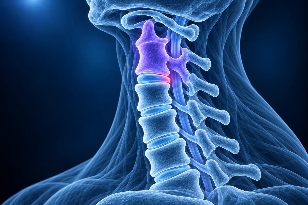
Klippel-Feil syndrome means two or more neck vertebrae were joined together before birth because they did not separate during early development. On an X-ray or CT, the fused bones share one block with little or no disc space between them. Many people have no symptoms; the main long-term concern is extra wear at the levels next to the fusion.
Common Symptoms
- No symptoms at all in many people, with the fusion found by chance on a neck X-ray or CT
- Reduced neck movement, most noticeable when turning the head to look over the shoulder
- A short-looking neck and a low hairline at the back of the head
- Neck pain or headaches that start in adolescence or adulthood as the levels next to the fusion wear
- Arm pain, tingling or weakness if a nerve root becomes pinched next to the fused block
- Hand clumsiness or unsteady walking if the spinal cord becomes compressed
When should someone with Klippel-Feil syndrome get urgent care?
People with congenitally fused neck vertebrae are at higher risk of nerve injury after neck trauma, because the moving levels next to the block take more of the force. After a fall, a car crash or a sports collision, get same-day assessment for neck pain, any tingling, numbness or weakness in the arms or legs, or a brief episode of burning or paralysis in the limbs, even if it has passed. Tell the emergency team about the fusion.
Also seek prompt medical review for hands getting clumsier, a change in walking or balance, new bladder problems, or neck pain with fever or a history of cancer. These can mean the spinal cord or a nerve is under pressure, or that something other than the fusion is going on. This page explains imaging findings; it does not replace an examination.
What is Klippel-Feil syndrome?
In the first weeks of pregnancy the spine forms as a column of segments that later separate into individual vertebrae with discs between them. In Klippel-Feil syndrome, some cervical segments fail to separate, so two or more vertebrae grow as one. It was first described in 1912 by Maurice Klippel and André Feil. The group of people who have it is very varied: some have a single fused pair and nothing else, others have long fused segments together with other birth differences. Researchers have linked it to genes that control how the spine segments, but most cases have no identified cause.
The textbook picture is a triad of a short neck, a low hairline at the back and limited neck movement, yet in a classic 1974 series of 50 patients fewer than half had all three. That is why the condition is now found mostly on imaging. Among 2,917 adults who had a neck CT in one emergency department, 17 had congenital fusion, about 1 in 172, far more than the older estimate of 1 in 40,000 births. All 17 had a single fused segment, most often at C5–C6 or C2–C3.
What does congenital fusion look like on a neck X-ray or CT?
- "Congenital fusion of C2–C3" or "block vertebra"
- Two vertebral bodies are joined into one taller block. Ask whether the fusion is single or multiple and whether it involves the top of the neck, because those details shape all later advice.
- "Wasp-waist sign"
- The fused block is narrower from front to back at the level where the disc should be, like a pinched waist. It points to a fusion present since birth rather than one caused by surgery or disease, so ask whether it was seen.
- "Rudimentary disc space" or "absent intervertebral disc"
- Only a faint line, or no gap at all, marks where the disc would be. Ask whether the discs above and below look normal for your age.
- "Fused facets and spinous processes"
- The small joints and bony tips at the back are joined as well as the bodies in front. This favours a congenital fusion and helps tell it apart from bone that has bridged later in life.
- "Adjacent segment degeneration" or "hypermobility at the adjacent level"
- The disc or joints next to the block show early wear or extra movement. Ask whether flexion-extension views or an MRI are needed to check stability and the space around the cord.
For other wording on neck films, see cervical spine X-ray explained.
Why does a congenital neck fusion matter later in life?
A fused block cannot bend, so the discs and joints just above and below it move more to make up the difference. Over the years those levels tend to wear faster. In a study that pooled 592 MRI scans from people treated for degenerative cervical myelopathy, congenital fusion was more common than in the general population, and cord compression tended to appear at the levels next to the fusions. In a separate follow-up of 28 children and teenagers, about a third developed neck-related symptoms, mostly neck pain, while arm or cord symptoms were more likely when several levels were fused.
That extra movement can turn into instability, where one vertebra shifts on the next when the neck bends. Fusions at the top of the neck, long fused segments, and a narrow canal next to the block add to the risk. The wear itself looks like ordinary cervical spondylosis, and severe narrowing next to a fusion behaves like cervical spinal stenosis, just at a younger age.
Which other conditions are checked with Klippel-Feil syndrome?
Klippel-Feil syndrome can come with differences elsewhere in the body, so the diagnosis is a reason to look further, especially in children. In the 1974 series of 50 patients, more than half had scoliosis and about a third had a kidney anomaly; 21 had Sprengel deformity, a shoulder blade that sits high, 15 had hearing impairment, 9 had mirror movements of the hands (synkinesia) and 7 had congenital heart disease. Many of these do not cause symptoms on their own.
Questions worth asking your doctor: has the rest of the spine been checked for scoliosis or other fused vertebrae, have the kidneys been looked at with ultrasound, has hearing been tested, and is a heart check needed? Adults diagnosed by chance may already have had some of these checks in childhood, so bring old records if you have them.
How is Klippel-Feil different from surgical fusion or DISH?
A surgical fusion usually shows plates, screws or a cage, a graft where the disc was, and no wasp waist. Your history makes it obvious in most cases, and cervical MRI after fusion explains what those scans look like. A congenital block has smooth bone throughout, no metal, and often a narrow waist with fused joints at the back.
DISH joins vertebrae in older adults with a thick, flowing band of new bone along the front of the spine, while the discs keep their height and the vertebral bodies keep their normal shape. Bone that bridges across very worn, collapsed discs in late life can also mimic a block. In contrast, Klippel-Feil fusion is present from childhood, joins the bodies themselves, and has little or no disc left between them.
Which scans are used for Klippel-Feil syndrome?
Plain X-rays of the neck are usually where the fusion is first seen. Flexion-extension views, taken with the head bent forward and back, show whether the levels next to the block move too much; the flexion-extension X-ray guide explains how they are read. CT maps the bony anatomy in three dimensions and is the test emergency teams use after an injury, guided by decision rules such as NEXUS and the Canadian C-spine Rule.
MRI without contrast is added when there are arm symptoms, cord symptoms or instability, in line with the ACR Appropriateness Criteria for cervical pain or radiculopathy, because it shows the discs, the nerve roots and the spinal cord, which X-ray and CT cannot. It can also show cord anomalies that sometimes accompany the fusion.
What we look for on Klippel-Feil imaging
- Which levels are fused, and whether there is one block, several separate blocks or one long segment
- A wasp-waist narrowing and fused facets or spinous processes that point to a congenital cause
- Disc wear, bone spurs or slipping at the levels directly above and below the block
- Canal width and any cord contact next to the fusion on MRI
- Involvement of the skull base and upper neck, and any curve in the cervical or upper thoracic spine
Can I play contact sports with Klippel-Feil syndrome?
It depends on the fusion pattern, and the decision belongs with a spine or sports medicine specialist who has seen your images. Sports guidance for collision activities rates congenital cervical fusions anywhere from no contraindication to an absolute contraindication. A single fused level with full neck movement and no instability, narrow canal or disc disease is generally viewed more permissively. Long fused segments, fusion involving the upper neck, instability on flexion-extension views, or a narrow canal next to the block usually lead to advice against collision sports.
Any episode of burning, numbness or brief weakness in the arms or legs after a hit means stopping play until an MRI and specialist review are done.
How is Klippel-Feil syndrome treated?
The fusion itself is not treated; there is nothing to reverse. Care focuses on the levels around it and on any associated conditions. People without symptoms are usually advised to avoid high-risk neck impacts and to get new arm, hand or walking symptoms checked early. Neck pain is managed in the usual ways, with exercise, physiotherapy and pain relief. Surgery is reserved for instability, nerve root compression that does not settle, or spinal cord compression, and then aims to decompress and stabilise the affected neighbouring level.
Can I see the fused vertebrae on my own scan?
Yes. A neck X-ray opens privately in the free X-ray viewer and a CT in the free CT viewer: on the side view, look for the block where a disc gap should be and for the narrow waist across it. Use the free MRI viewer to check the discs and cord next to the fusion. For a plain-language explanation of your images and report, upload them for an AI review. It explains what the images show; it is not a diagnosis. For wording about the levels beside a congenital fusion, see the cervical MRI report glossary.
Frequently Asked Questions
Is Klippel-Feil syndrome rare?
It is more common than once thought. Older estimates were about 1 in 40,000 births, but a CT study of 2,917 adults found congenital neck fusion in about 1 in 172, mostly a single fused level without symptoms.
Is Klippel-Feil syndrome hereditary?
Some families show an inherited pattern and several genes have been linked to it, but most people have no affected relatives and no single cause is found. A clinical genetics referral can help if several family members are affected.
Will Klippel-Feil syndrome get worse with age?
The fused block does not change, but the levels next to it can wear faster than the rest of the neck. Many people stay symptom-free; new arm symptoms, hand clumsiness or walking changes are the reasons to get re-checked.
Can a congenital fusion be mistaken for an old fracture or surgery?
Sometimes on a single X-ray. A wasp-waist shape, fused joints at the back and no hardware favour a congenital fusion; CT settles most doubtful cases.
Does Klippel-Feil syndrome need an MRI?
Not always. A fusion found by chance with no symptoms is often followed with X-rays. MRI is used when there are arm or cord symptoms, signs of instability, or before planning surgery.
Sources
- Menger RP, Rayi A, Notarianni C. Klippel Feil Syndrome. StatPearls. Treasure Island (FL): StatPearls Publishing.
- Hensinger RN, Lang JE, MacEwen GD. Klippel-Feil syndrome; a constellation of associated anomalies. J Bone Joint Surg Am. 1974;56(6):1246-1253.
- Gruber J, Saleh A, Bakhsh W, Rubery PT, Mesfin A. The Prevalence of Klippel-Feil Syndrome: A Computed Tomography-Based Analysis of 2,917 Patients. Spine Deform. 2018;6(4):448-453.
- Samartzis DD, Herman J, Lubicky JP, Shen FH. Classification of congenitally fused cervical patterns in Klippel-Feil patients: epidemiology and role in the development of cervical spine-related symptoms. Spine (Phila Pa 1976). 2006;31(21):E798-E804.
- Nouri A, Martin AR, Lange SF, Kotter MRN, Mikulis DJ, Fehlings MG. Congenital Cervical Fusion as a Risk Factor for Development of Degenerative Cervical Myelopathy. World Neurosurg. 2017;100:531-539.
- Torg JS, Ramsey-Emrhein JA. Management guidelines for participation in collision activities with congenital, developmental, or postinjury lesions involving the cervical spine. Clin J Sport Med. 1997;7(4):273-291.
- Ridley LJ, Han J, Ridley WE, Xiang H. Wasp waist sign: Congenital vertebral fusion. J Med Imaging Radiat Oncol. 2018;62(Suppl 1):166-167.
- Expert Panel on Neurological Imaging; Eldaya RW, Parsons MS, Hutchins TA, et al. ACR Appropriateness Criteria Cervical Pain or Cervical Radiculopathy: 2024 Update. J Am Coll Radiol. 2025;22(5S):S136-S162.
Understand your imaging before your next appointment
MRI, X-ray or CT from a CD, folder or ZIP. A PDF report or photos of the films work too.
Free to open and preview, no account needed. An optional AI report from $10 explains your images in plain language, with questions for your doctor.
Your raw files stay on this device. Only rendered images and minimal context are sent when you request an AI report.
Medical Disclaimer: This page is for informational and educational purposes only. It does not constitute medical advice, diagnosis, or treatment. AI-generated analysis may contain errors. Always consult a qualified healthcare professional for medical decisions. Full Disclaimer