
Neck Herniated Disc Recovery Timeline: Phases and Re-Scans
How a herniated disc in the neck recovers phase by phase, how often herniations shrink on their own, when a repeat MRI helps, and which signs change the plan.
Key Takeaways
- Substantial improvement usually comes within four to six months of a cervical disc herniation
- About 83% of patients recovered completely within 24 to 36 months in a systematic review
- Extruded and migrated fragments are the herniations most likely to shrink on their own
- A repeat MRI helps when symptoms worsen or treatment is being planned, not to confirm recovery
- Worsening weakness, clumsy hands, an unsteady walk or bladder change need prompt assessment
Most people with a herniated disc in the neck and a pinched nerve improve a great deal within four to six months, usually without surgery. In a systematic review of how these herniations behave, about 83% of patients had recovered completely within 24 to 36 months. Arm pain tends to ease first; tingling and weakness can lag behind.
That long tail is why the recovery feels slow even when it is going well. This guide splits the course into phases, explains how often the disc fragment itself shrinks, sets out when a repeat MRI earns its place and when it does not, and lists the symptoms that change the plan on any day. It is general education about a common pattern, not a forecast for your own neck; the clinician who examines you sets the pace.
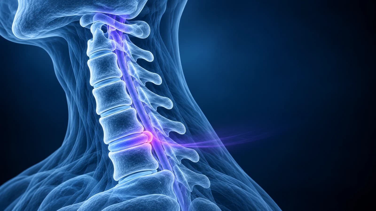
Which symptoms stop the waiting and need prompt care?
The timeline below assumes a nerve-root problem that is stable or improving. Contact a clinician the same day, or go to an emergency department if the change is sudden or severe, when any of these appear:
- Weakness in the arm or hand that is getting worse from one week to the next, such as a dropping grip
- Clumsy hands, trouble with buttons or handwriting, or a new unsteady, wide-based walk
- Numbness or tingling spreading to both arms, or into the legs
- New trouble starting or controlling urination, or loss of bowel control
- Fever, unexplained weight loss, a history of cancer, or pain that is relentless at night and at rest
The middle three point toward pressure on the spinal cord rather than on a single nerve root, which is explained in cervical myelopathy. Cord compression is treated on a different, faster clock than an arm-pain episode.
How long does a herniated disc in the neck take to heal?
There is no fixed calendar, and the phases below overlap. They describe a typical cervical disc herniation that irritates one nerve root without pressing on the cord. The North American Spine Society guideline puts it plainly: for most people the symptoms are likely to be self-limited and to resolve over a variable length of time.
- First weeks: the hardest stretch. Pain often runs from the neck over the shoulder blade and down the arm, sleep is broken and certain head positions make it flare. In a population study from Rochester, Minnesota, people had lived with symptoms for a median of 15 days by the time the diagnosis was made.
- Weeks 6 to 12: for many, the arm pain is noticeably lower and nights are easier, while pins and needles in the fingers persist. This is the usual point at which a clinician reviews whether conservative care is working.
- Months 3 to 6: the review by Wong and colleagues found that substantial improvement tends to happen within the first four to six months after onset, even though most patients start with intense pain and moderate disability.
- Beyond 6 months: what remains is often a patch of numbness, mild tiredness of the arm muscles or occasional twinges. Complete recovery took 24 to 36 months in about 83% of patients in that review, and in the Rochester study 90% were free of symptoms or only mildly limited at a median follow-up of 4.9 years.
Which nerve is pinched shapes what you feel along the way. A herniation at C5–C6 usually affects the C6 root and the thumb side of the hand, while one at C6–C7 affects the C7 root and the middle finger; the patterns are mapped in C5–C6 and C6–C7 disc herniation explained.
Why does the pain ease before the numbness and weakness?
Much of the early pain is thought to come from chemical inflammation around the nerve root, not only from the squeeze. As that inflammation settles, pain can drop even while the disc material is still in place. Sensation and muscle strength depend on the nerve fibres themselves conducting normally again, and nerve tissue recovers slowly, so a numb fingertip or a weaker triceps can outlast the pain by months.
The practical point is the direction of travel. Weakness that is stable or slowly improving fits the expected course. Weakness that is getting worse is one of the clearest reasons to bring a review forward, whatever the calendar says.
Does a herniated disc in the neck shrink on its own?
Often, yes. Disc material that has pushed out into the space around the nerve is treated by the body as foreign tissue: it draws in blood vessels and immune cells that break it down. In a Japanese study of 38 patients with cervical disc herniation who had repeated MRI scans, the herniated material became smaller in 15, or 40%. Fragments that had extruded beyond the ligament behind the disc, and those that had travelled up or down or out to the side, were the ones most likely to shrink.
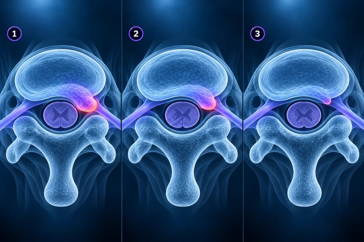
A 2021 literature review gathered 76 published cases of cervical herniations that regressed on MRI, most of them at C5–C6 or C6–C7, with regression documented on MRI after an average of about nine months. Case collections like this show that it happens, not how often, so the most useful pooled numbers still come from the lower back.
For lumbar herniations, a systematic review estimated spontaneous regression in 96% of sequestered fragments, 70% of extrusions, 41% of protrusions and 13% of bulges. The same ranking, with the biggest, most displaced herniations shrinking most, matches the cervical studies above. The lower-back version of this story is told in can a herniated disc heal on its own. What the four shape words mean on a neck report is set out in disc bulge vs herniation in the neck.
Do you need a repeat MRI to check that the disc has healed?
Usually not while you are getting better. Symptoms and the examination are the main measures of recovery, and the picture tends to trail the person: pain can fade months before a scan would show the fragment smaller, and some people feel well with a herniation that looks unchanged. A routine follow-up scan in someone who is improving rarely changes what happens next.
Imaging, or a repeat of it, becomes useful in a few situations:
- New or increasing radiculopathy, such as spreading numbness or a new weak muscle, where the ACR Appropriateness Criteria rate MRI without contrast as usually appropriate
- Arm symptoms that have not responded to a course of conservative care in someone who may be a candidate for an injection or surgery; the NASS guideline suggests MRI to confirm a compressive lesion that matches the symptoms
- Any sign that the spinal cord, not just a nerve root, is involved
- Fever, a cancer history or other red flags, where MRI with and without contrast may be chosen
If a repeat scan is done, ask whether it will be compared directly with the first one. You can open both studies yourself in the free MRI viewer, which runs in your browser without uploading the files, and scroll to the same disc level on the axial slices.
What do follow-up MRI reports say about a healing disc?
- “Interval decrease in size of the right paracentral disc extrusion at C6–C7” — the fragment is smaller than on the previous scan, the pattern that matches resorption; ask whether the nerve root now looks free
- “Residual small protrusion” — some disc tissue remains beyond the normal margin; ask whether it still touches a nerve root on the painful side
- “Resolution of previously seen foraminal narrowing” — the opening where the nerve exits is no longer crowded by disc material
- “Stable appearance compared with the prior study” — no change in size; ask how that fits with your symptoms, because stable images and improving symptoms often go together
- “New cord signal abnormality” or “increased cord compression” — a change at the spinal cord itself, which needs a prompt conversation with the treating team
Can a herniated disc in the neck come back?
Yes, a second episode is common. In the Rochester study, which followed 561 people with cervical radiculopathy for a median of 4.9 years, symptoms recurred in 31.7% and 26% had an operation at some point. Not every episode is a soft herniation: a confirmed disc protrusion was the cause in only 21.9% of that group, while most cases involved spondylosis, the bony and disc wear of ageing, and a narrowed nerve exit can flare without any new disc material.
When the arm symptoms come from a bony spur rather than soft disc, the outlook and the treatment can differ, as explained in cervical radiculopathy. A recurrence with the same pattern as the first episode often follows a similar course.
When does surgery come into the timeline?
For an arm-pain problem without cord compression, surgery is usually discussed when pain stays disabling after a fair trial of conservative care, or when weakness is significant or worsening. The NASS guideline suggests that surgery gives faster relief than non-operative treatment; in the small randomised trial it reviewed, outcomes after surgery and after collar or physiotherapy were similar at one year. Waiting is therefore a reasonable choice for many people, and speed of relief is a reasonable reason not to wait for others.
Cord compression with symptoms changes the arithmetic, because lost cord function may not come back; the warning signs on imaging are covered in cervical myelopathy warning signs on MRI. If an operation is on the table, our list of questions to ask before neck surgery helps you prepare.
Frequently Asked Questions
How long does a pinched nerve from a neck disc last?
The worst pain usually eases over weeks, and most people improve substantially within four to six months. Residual tingling or mild weakness can take longer; in a systematic review, complete recovery took 24 to 36 months in about 83% of patients. A clinician should reassess anyone whose symptoms are getting worse rather than better.
Will my herniated disc be gone on the next MRI?
It may be smaller, but it may also look unchanged while you feel much better. In one cervical study, herniations shrank in 40% of patients who had repeat scans, most often when the fragment had extruded or migrated. The follow-up scan is judged against your symptoms, not on its own.
Is a bigger herniation worse for recovery?
Not necessarily. Large extruded or sequestered fragments are the ones most often resorbed, while a small contained protrusion shrinks less often. Size matters most when the disc presses on the spinal cord, because cord compression with symptoms is managed differently from a pinched nerve root.
Can I make the disc heal faster?
Treatment works mainly on the symptoms rather than on the disc fragment itself. Staying active within comfort, pain relief that suits you, guided exercise and, for some people, a targeted injection aim to control symptoms while the natural course runs. Which of these fits is a decision for you and your clinician.
Can an AI explanation tell me how long my recovery will take?
No. It can describe what your MRI shows, translate the report wording and point out what changed between two reports, which helps you prepare questions. It is not a diagnosis and cannot examine your arm strength or reflexes, so the outlook for your own neck stays with your treating clinician.
Sources
- Wong JJ, Côté P, Quesnele JJ, Stern PJ, Mior SA. The course and prognostic factors of symptomatic cervical disc herniation with radiculopathy: a systematic review of the literature. Spine J. 2014;14(8):1781-1789.
- Radhakrishnan K, Litchy WJ, O'Fallon WM, Kurland LT. Epidemiology of cervical radiculopathy. A population-based study from Rochester, Minnesota, 1976 through 1990. Brain. 1994;117(Pt 2):325-335.
- Mochida K, Komori H, Okawa A, et al. Regression of cervical disc herniation observed on magnetic resonance images. Spine (Phila Pa 1976). 1998;23(9):990-995.
- Sharma AK, Gandhoke CS, Syal SK. Spontaneous regression of herniated cervical disc: a case report and literature review. Surg Neurol Int. 2021;12:141.
- Chiu CC, Chuang TY, Chang KH, et al. The probability of spontaneous regression of lumbar herniated disc: a systematic review. Clin Rehabil. 2015;29(2):184-195.
- Bono CM, Ghiselli G, Gilbert TJ, et al. An evidence-based clinical guideline for the diagnosis and treatment of cervical radiculopathy from degenerative disorders. Spine J. 2011;11(1):64-72.
- Expert Panel on Neurological Imaging; McDonald MA, Kirsch CFE, et al. ACR Appropriateness Criteria Cervical Neck Pain or Cervical Radiculopathy. J Am Coll Radiol. 2019;16(5S):S57-S76.
Related Articles
Why C5-C6 and C6-C7 are the neck discs that herniate most, which nerve root each one pinches, how the arm symptoms differ, and what protrusion, extrusion and foraminal mean on your MRI.
Bulge, protrusion, extrusion and sequestration on a neck MRI in plain words: how the shapes differ, why bulges are common without pain, and when it matters.
Questions to ask before neck surgery: does the MRI match your symptoms, ACDF vs disc replacement vs foraminotomy, recovery, adjacent levels and the real risks.
Related Conditions
Understand your imaging before your next appointment
MRI, X-ray or CT from a CD, folder or ZIP. A PDF report or photos of the films work too.
Free to open and preview, no account needed. An optional AI report from $10 explains your images in plain language, with questions for your doctor.
Your raw files stay on this device. Only rendered images and minimal context are sent when you request an AI report.
Medical Disclaimer: This page is for informational and educational purposes only. It does not constitute medical advice, diagnosis, or treatment. AI-generated analysis may contain errors. Always consult a qualified healthcare professional for medical decisions. Full Disclaimer