
Disc Bulge vs Herniation in the Neck: What Your MRI Means
Bulge, protrusion, extrusion and sequestration on a neck MRI in plain words: how the shapes differ, why bulges are common without pain, and when it matters.
Key Takeaways
- A bulge is a broad overhang of more than a quarter of the disc rim; a herniation is focal
- Protrusion has a base wider than its reach; extrusion is wider at the tip; sequestration is a free piece
- Bulges appeared in 87.6% of 1,211 people without neck symptoms, even in their twenties
- What matters most is contact with the nerve root on the painful side or with the spinal cord
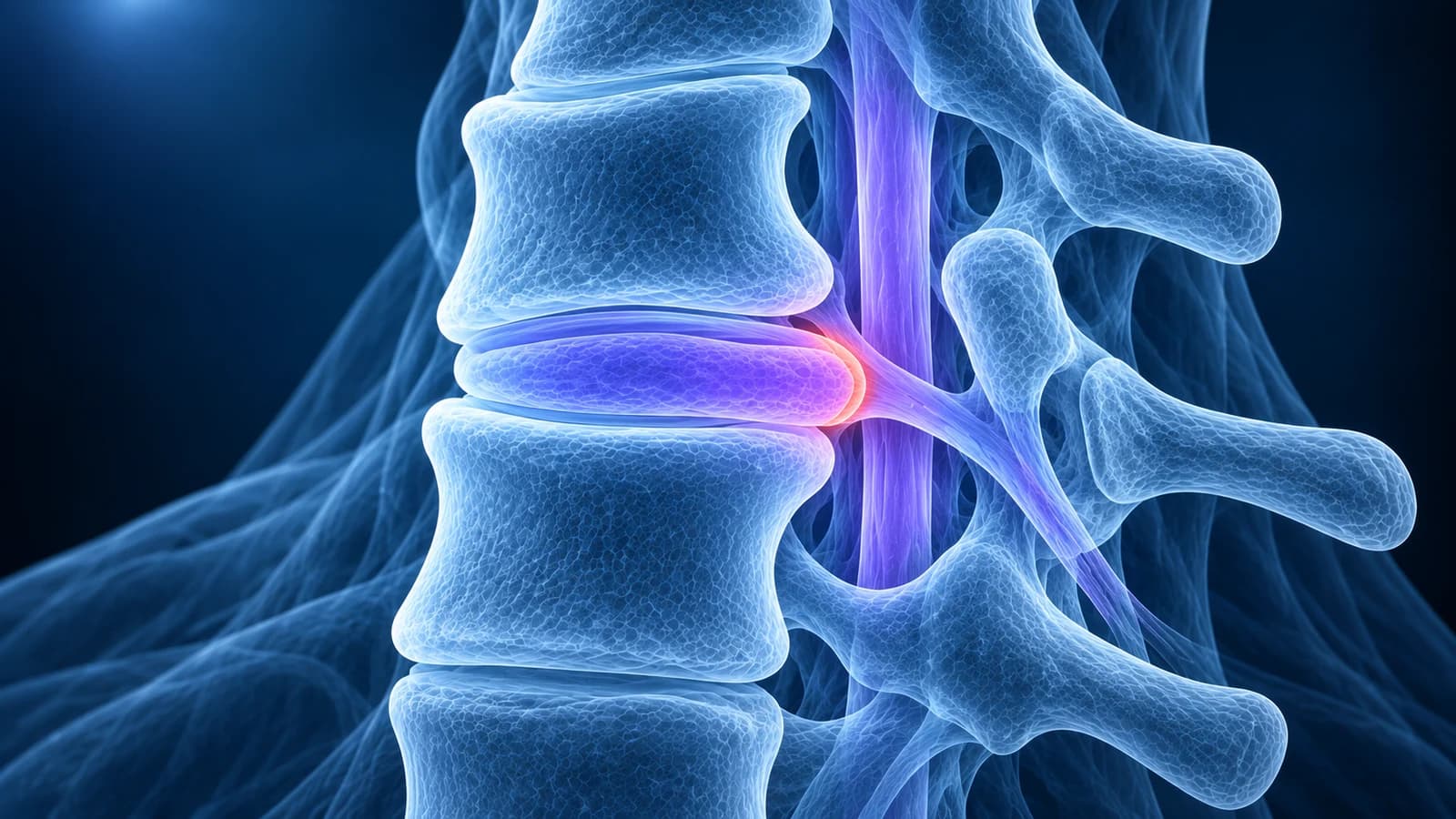
A disc bulge in the neck is a broad, shallow overhang of the whole disc rim past the edge of the vertebra, usually an age-related change; a herniation is a focal piece of disc pushed out in one spot. Bulges were found in 87.6% of 1,211 people with no neck symptoms, so the shape matters less than whether the disc touches a nerve root or the cord.
Radiologists use a shared vocabulary for these shapes, written up by the North American Spine Society and two radiology societies in 2014. It was drafted for the lower back, but the same definitions are used to describe the cervical discs. This guide translates that vocabulary for the neck, shows what each term looks like and explains the words around it that decide whether a finding is likely to matter. It is educational and does not replace your radiologist’s report or your clinician’s examination.
What is the difference between a disc bulge and a disc herniation?
The difference is how much of the disc’s outline is involved. Picture the disc from above as a round cushion sitting on the vertebra. In a bulge, the rim of the cushion overhangs the bone around a large part of its edge, more than a quarter of the circle, and usually by only a few millimetres. In a herniation, disc material is displaced in one limited spot, less than a quarter of the circle, like a thumb pressed out of the side of a pillow.
The 2014 nomenclature is explicit that a bulge is not a type of herniation. A bulge reflects the disc losing height and spreading as it dries out with age, or the bones settling around it. A herniation means material from inside the disc has moved beyond its normal space, usually through a fissure in the tough outer ring. Herniations are then split by shape into protrusion and extrusion, and an extrusion that has broken free becomes a sequestration.
What do bulge, protrusion, extrusion and sequestration look like?
Radiologists judge the shape on the axial slice, the one that cuts straight across the disc, and confirm it on the sagittal slice that runs the length of the neck. The test that separates the two herniation types is simple: compare the width of the displaced material where it joins the disc (its base) with how far it reaches out.
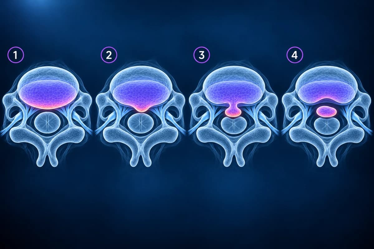
- Bulge: an even overhang around a broad stretch of the rim. Reports may add “broad-based” or “circumferential”, and an “asymmetric bulge” leans more to one side.
- Protrusion: a focal herniation that stays broad at its base and short in reach. Many protrusions in the neck are small and sit in the midline, where the canal is widest.
- Extrusion: in at least one view the displaced piece is wider than its connection to the disc, or it has slid up or down behind a vertebral body. Extrusions tend to be the cause of a sudden, sharp attack of arm pain.
- Sequestration: an extrusion that has lost all contact with the parent disc, sometimes called a free fragment. The report should say where it now lies, because a surgeon would need to find it.
In the neck these shapes are often blended with bone. A disc bulge merged with spurs from the vertebral corners is reported as a disc osteophyte complex, explained in disc osteophyte complex explained. That matters because bone does not shrink the way soft disc material can.
Why do so many people have disc bulges in the neck without pain?
Because bulging is part of how discs age. A Japanese study scanned 1,211 volunteers aged 20 to 70 who had no neck or arm symptoms. Disc bulging was found in 87.6% of them, and it was already present in 73.3% of men and 78.0% of women in their twenties. Bulges became more frequent, larger and spread over more levels with each decade. By contrast, only 5.3% had a disc pressing on the spinal cord and 2.3% had a bright signal change inside the cord, mostly after the age of 50.
An older American study made the same point with true herniations. Neuroradiologists reading scans of 63 people without symptoms, mixed blindly with scans of patients, called 19% of them abnormal: 14% under the age of 40 and 28% over it. A worn or narrowed disc appeared in 25% of those under 40 and in almost 60% of those over 40. The authors warned against basing an operation on a scan finding that does not match the patient’s signs.
So a report listing “disc bulges C3-C4 through C6-C7” is describing the common picture of cervical spondylosis. It does not, by itself, explain neck pain, and it is not a reason for surgery.
When does the wording on a neck MRI report actually matter?
The shape word is less important than the words that follow it. Two questions decide whether a cervical disc finding is likely to be relevant: is it touching or squeezing a nerve root on the side of your symptoms, and is it touching or squeezing the spinal cord? Read the rest of the sentence for these phrases.
- “Contacting”, “abutting” or “displacing the exiting right C7 nerve root”: the disc reaches a specific nerve. If that nerve matches the arm where you feel pain, tingling or weakness, the finding probably explains your cervical radiculopathy. Ask whether the side and level match your examination.
- “Narrowing the left neural foramen”: the side exit the nerve passes through is tighter. In the neck this is often due to bone spurs as well as disc. Ask how severe the narrowing is graded.
- “Effacing the ventral CSF” or “indenting the thecal sac”: the disc pushes into the fluid in front of the cord but does not touch the cord. Usually not urgent on its own.
- “Contacting”, “flattening” or “compressing the spinal cord”: the disc reaches the cord. A widely used grading system calls a deformed cord grade 2 and a bright signal change inside the cord grade 3. Ask your clinician to check for signs of cervical myelopathy such as clumsy hands or a change in walking.
- “Migrated superiorly” or “inferiorly”: an extruded or sequestered piece has moved above or below the disc. Ask which level it now sits behind, because the symptoms can follow that level.
A small protrusion that compresses the cord matters more than a large bulge that touches nothing, and an extrusion beside a nerve root on the painful side matters more than three bulges in the midline. More report lines are translated in the cervical MRI report phrases guide.
What do “central”, “paracentral” and “foraminal” mean?
These words say where around the back of the disc the material points, and they often predict the symptoms. A central herniation points straight back toward the middle of the cord. A paracentral (or subarticular) one sits just off the midline, where it can press on the edge of the cord and the start of a nerve root. A foraminal herniation pushes sideways into the exit tunnel and tends to pinch a single root, which is the typical cause of pain running down one arm. A herniation that reaches beyond the tunnel is called extraforaminal.
Because of the way nerves are numbered in the neck, a foraminal herniation at C5-C6 affects the C6 root and one at C6-C7 affects the C7 root. Those two levels and their typical arm patterns are compared in C5-C6 and C6-C7 disc herniation explained.
Is an extrusion worse than a protrusion?
Not automatically. An extrusion is a larger displacement and is more often linked with a sudden attack of arm pain, but the shape word does not predict how you will recover. A review of the course of symptomatic cervical disc herniation found that most people improve substantially within the first four to six months, and most recover. Soft extruded material can also shrink as the body clears it, which is one reason surgery is usually reserved for weakness that is getting worse, cord compression with myelopathy, or pain that stays severe despite treatment.
The comparison also depends on the canal. The cervical canal is narrow in some people from birth, and a modest protrusion in a tight canal can touch the cord where a larger one would not. If your report also mentions cervical spinal stenosis, ask how much room is left around the cord at the level of the disc finding.
Does a disc bulge in the neck mean the same as one in the lower back?
The words are the same, but the stakes differ. In the lower back the canal holds loose nerve roots, so even a big herniation usually affects nerves to the leg. In the neck the canal holds the spinal cord itself, so a disc that reaches the middle of the canal can affect the hands, the legs and balance. That is why neck reports always state what the disc does to the cord. If your question is about a lumbar disc, see herniated disc in the lower back.
Which scan shows whether a neck disc is bulging or herniated?
Only MRI shows the disc itself well enough to name its shape. X-rays show the bones, the spacing between them and spurs, but not the disc material or the cord. CT shows bony spurs and calcified disc sharply but separates soft disc from the nerves less clearly. The American College of Radiology rates MRI of the cervical spine without contrast as the usual test for long-standing neck pain with arm symptoms, while for new arm pain without warning signs it rates no test as routinely needed at first. When MRI is not possible, the comparison in CT vs MRI for cervical radiculopathy explains what CT can still answer.
When should a disc finding be checked urgently?
Whatever word your report uses, seek same-day medical care if you have arm or hand weakness that is getting worse, clumsy hands (dropping things, trouble with buttons or writing), a new change in walking or balance, new bladder or bowel problems, fever with neck pain, a history of cancer, or neck pain after a significant fall or crash. These can be signs of cord compression, infection or injury, and they take priority over any reading of the scan.
How can I see the disc on my own MRI?
Open the DICOM files from your disc in the free MRI viewer, which reads them in your browser without uploading the originals. On the sagittal T2 series, count down from C2 to the level your report names. Then switch to the axial series at that disc: the cord is the round grey shape in the middle of the bright fluid, and the disc is the darker grey band in front of it. A bulge looks like a smooth even rim; a herniation looks like a localised bump, and an extrusion like a knob on a stalk.
Frequently Asked Questions
Is a broad-based disc bulge the same as a herniated disc?
No. A broad-based bulge involves a large part of the disc rim and is usually an age-related change. A herniation is a focal displacement of disc material. Some reports use “broad-based protrusion” for a herniation covering a quarter to half of the rim, which is different again, so ask your clinician which they mean.
Can a cervical disc bulge cause arm pain?
It can when it combines with bone spurs to narrow a nerve exit, especially in an already tight foramen. A bulge that touches no nerve and leaves the canal open is unlikely to explain arm symptoms, and the cause may lie elsewhere.
Can a neck disc herniation go back in?
The material does not usually slip back, but soft herniated tissue can shrink over months as the body clears it, and symptoms often settle even when the scan changes little. Repeat imaging is generally only needed if the plan would change.
What does “annular fissure” or “annular tear” mean next to a bulge?
It is a split in the outer ring of the disc, often seen as a small bright spot at its back edge. The 2014 nomenclature prefers “fissure” because the word “tear” wrongly suggests an injury. Fissures are common with age and are not a herniation by themselves.
Does a disc bulge need surgery?
Almost never on its own. Surgery for a cervical disc is considered when a herniation or disc osteophyte complex matches progressive weakness, cord compression with myelopathy, or severe pain that has not settled with treatment.
Sources
- Fardon DF, Williams AL, Dohring EJ, et al. Lumbar disc nomenclature: version 2.0. Recommendations of the combined task forces of the North American Spine Society, the American Society of Spine Radiology and the American Society of Neuroradiology. Spine J. 2014;14(11):2525-2545.
- Nakashima H, Yukawa Y, Suda K, et al. Abnormal findings on magnetic resonance images of the cervical spines in 1211 asymptomatic subjects. Spine (Phila Pa 1976). 2015;40(6):392-398.
- Boden SD, McCowin PR, Davis DO, et al. Abnormal magnetic-resonance scans of the cervical spine in asymptomatic subjects. A prospective investigation. J Bone Joint Surg Am. 1990;72(8):1178-1184.
- Kang Y, Lee JW, Koh YH, et al. New MRI grading system for the cervical canal stenosis. AJR Am J Roentgenol. 2011;197(1):W134-W140.
- Wong JJ, Côté P, Quesnele JJ, Stern PJ, Mior SA. The course and prognostic factors of symptomatic cervical disc herniation with radiculopathy: a systematic review of the literature. Spine J. 2014;14(8):1781-1789.
- Expert Panel on Neurological Imaging; McDonald MA, Kirsch CFE, Amin BY, et al. ACR Appropriateness Criteria Cervical Neck Pain or Cervical Radiculopathy. J Am Coll Radiol. 2019;16(5S):S57-S76.
Related Articles
Why C5-C6 and C6-C7 are the neck discs that herniate most, which nerve root each one pinches, how the arm symptoms differ, and what protrusion, extrusion and foraminal mean on your MRI.
What your neck MRI report means: uncovertebral hypertrophy, foraminal narrowing grades, effacement of the CSF, cord flattening, T2 hyperintense cord signal, Modic changes and more, each explained in one or two plain sentences.
A plain-language guide to "disc osteophyte complex" on a cervical MRI: what the ridge is made of, how it differs from a herniated disc, and what effacing the thecal sac, cord contact and cord compression mean.
Related Conditions
Understand your imaging before your next appointment
MRI, X-ray or CT from a CD, folder or ZIP. A PDF report or photos of the films work too.
Free to open and preview, no account needed. An optional AI report from $10 explains your images in plain language, with questions for your doctor.
Your raw files stay on this device. Only rendered images and minimal context are sent when you request an AI report.
Medical Disclaimer: This page is for informational and educational purposes only. It does not constitute medical advice, diagnosis, or treatment. AI-generated analysis may contain errors. Always consult a qualified healthcare professional for medical decisions. Full Disclaimer